

Introduction
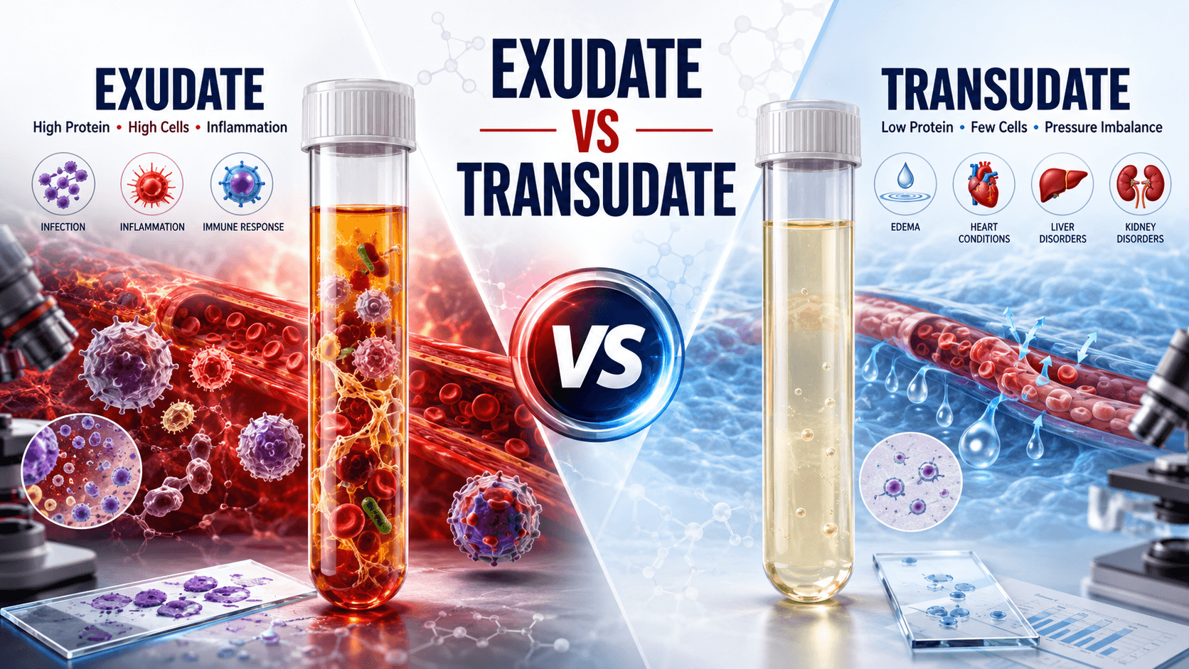
When fluid accumulates in the pleural, peritoneal, or pericardial cavities of a canine, feline, equine, or bovine patient, clinicians face a diagnostic critical point. Fluid accumulation is rarely a primary disease; rather, it is a liquid manifestation of systemic hemodynamic failure, microvascular disruption, severe metabolic compromise, or localized inflammation. Understanding exudate vs transudate veterinary pathology is fundamental to unravelling these underlying systemic disorders. Obtaining a cavity fluid sample via thoracocentesis or abdominocentesis is one of the most accessible, rapid, and high-yield diagnostic procedures available in veterinary practice. However, retrieving the fluid is only the first step. The true diagnostic value lies in evaluating its physical, biochemical, and cytological characteristics through a comprehensive veterinary effusion diagnosis guide. By applying established protocols—including Light’s criteria veterinary fluid analysis and assessing parameters such as the pleural fluid protein threshold canine clinicians—practitioners can definitively differentiate between exudative and transudative effusions.
Mastering canine peritoneal fluid interpretation and feline ascites exudate transudate profiles enables field practitioners to make swift, life-saving clinical decisions, guides students through complex pathophysiological pathways, and offers educators a clear, structured framework for veterinary clinical pathology body fluids instruction.
Effusion analysis is one of the most immediate, cost-effective, and high-yield diagnostic tools available in veterinary medicine. When fluid accumulates in the pleural, peritoneal, or pericardial cavities, it is not merely a symptom of disease—it is a liquid biopsy waiting to be interpreted. Differentiating between exudative and transudative effusions provides crucial insight into the underlying pathophysiological mechanisms, directly guiding emergency management, therapeutic protocols, and prognosis.
1. Pathophysiology of Fluid Accumulation.
In a healthy patient, cavity fluids (pleural, peritoneal, pericardial) exist in microscopic amounts to lubricate visceral membranes. Fluid movement between blood vessels and body cavities is governed by Starling’s forces:
Hydrostatic Pressure: Pushes fluid out of capillaries.
Colloid Osmotic (Oncotic) Pressure: Retains fluid inside capillaries, primarily driven by plasma albumin.
Vascular Permeability: Controls the passage of proteins and inflammatory cells across the endothelium.
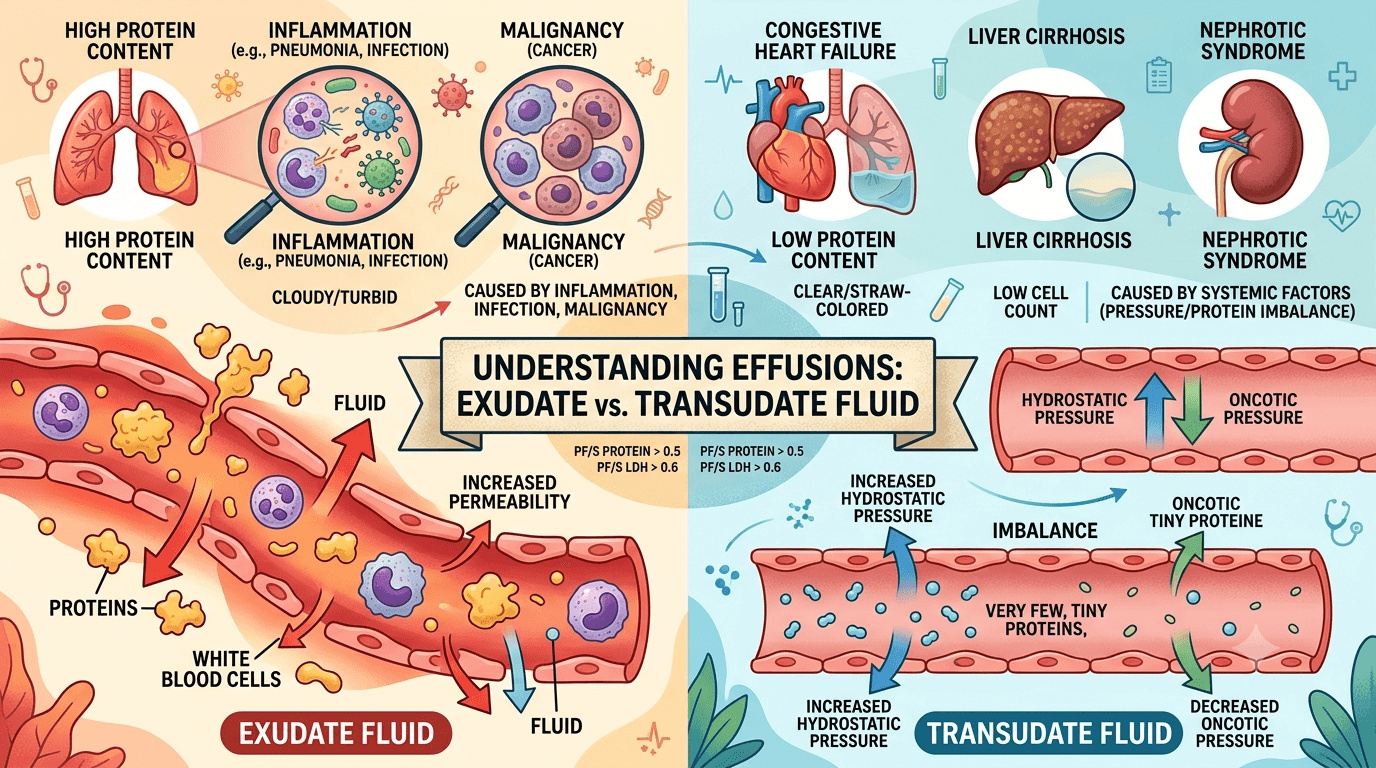
Disruption of these mechanisms leads to two distinct types of effusions:
Transudates
It result from systemic hemodynamic imbalances—either elevated hydrostatic pressure (e.g., congestive heart failure) or reduced plasma oncotic pressure (e.g., severe hypoalbuminemia). The vascular endothelium remains intact; thus, protein and cellular leakages are minimal.
Increased Hydrostatic Pressure: Venous congestion (e.g., Right-Sided Congestive Heart Failure, Portal Hypertension) forces protein-poor fluid into body cavities.
Decreased Plasma Oncotic Pressure: Severe hypoalbuminemia (serum albumin < 1.5\g/dL) due to hepatic insufficiency, Protein-Losing Nephropathy (PLN), or Protein-Losing Enteropathy (PLE) prevents the retention of intravascular fluid.
Exudates
stem from localized active inflammation, endothelial injury, or malignancy. Cytokines and inflammatory mediators induce microvascular hyperpermeability, allowing large plasma proteins, enzymes, and white blood cells to cross into the cavitary space.
Exudates result from active inflammatory processes, tissue injury, or neoplasia. Inflammatory cytokines (e.g., TNF-, IL-1, histamine) cause vascular endothelial cells to contract, dramatically increasing vascular permeability.
Large plasma proteins, enzymes, and inflammatory cells leak directly into the cavitary space.
2. Diagnostic Comparison: Exudate vs. Transudate
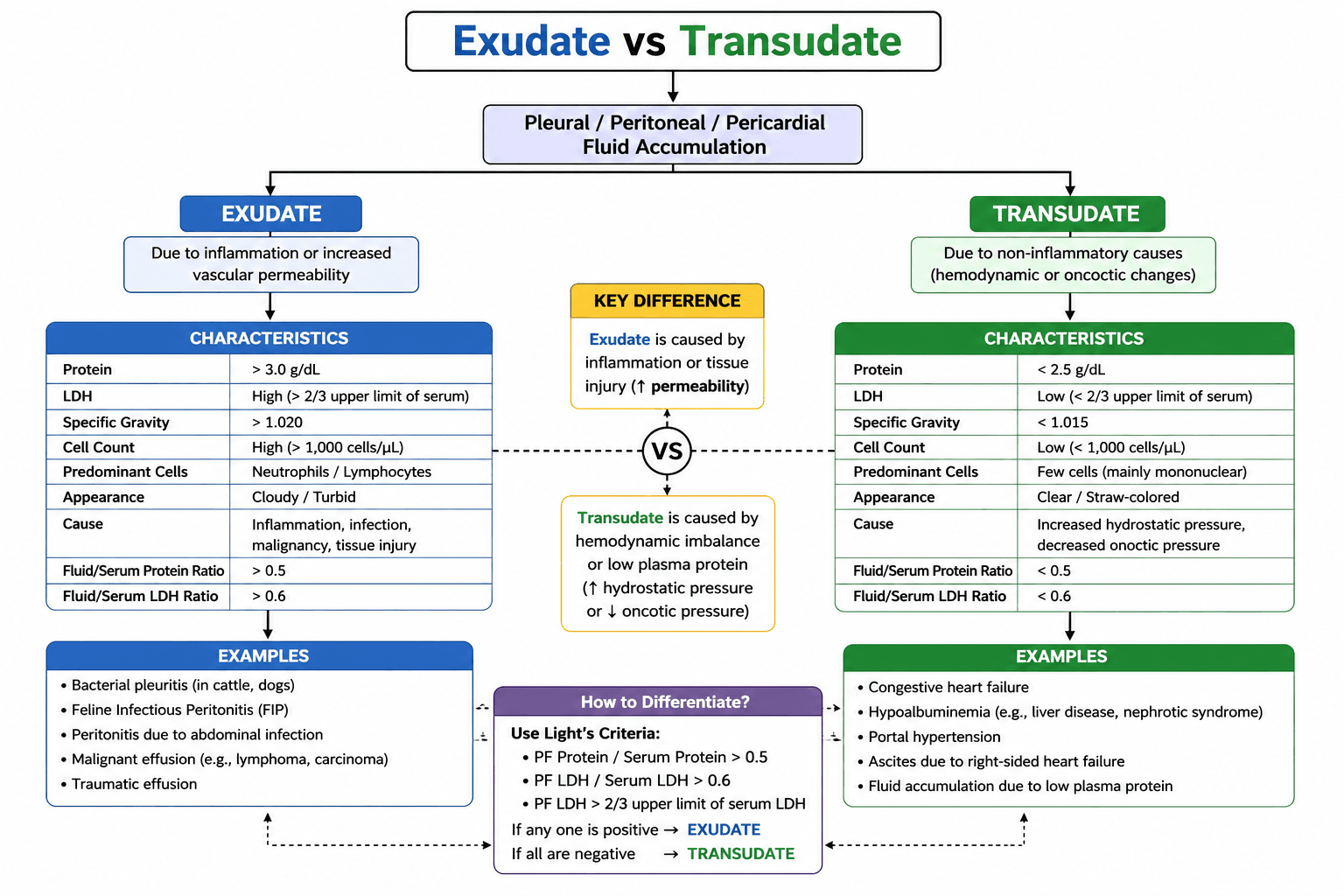
Accurate clinical diagnosis relies on quantitative fluid analysis. The table below details the physical, biochemical, and cytological benchmarks used to differentiate these fluids:
3. Light’s Criteria in Veterinary Medicine Transudate
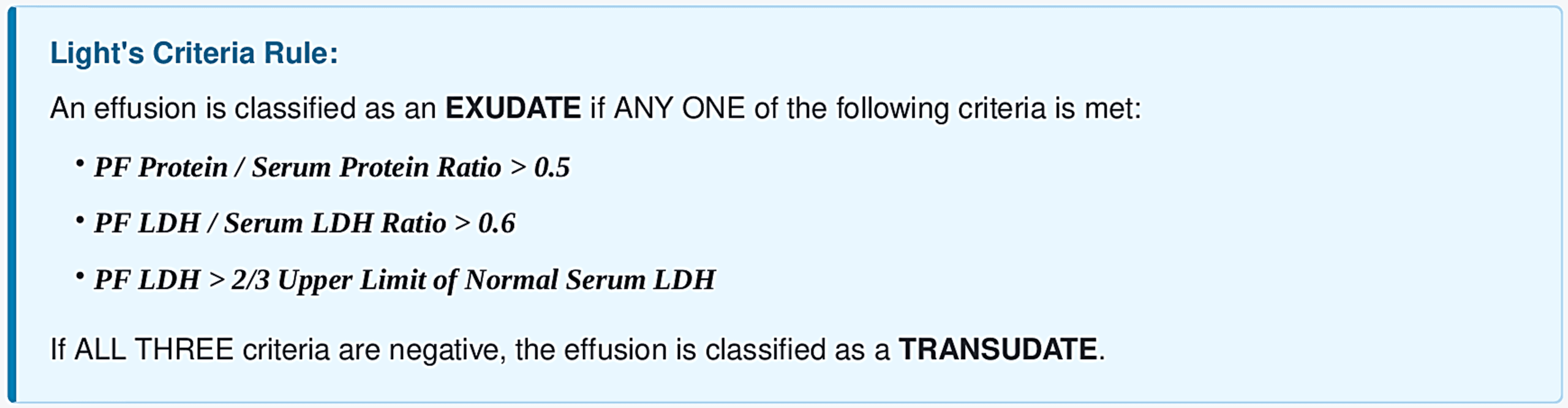
While originally established in human medicine, Light’s Criteria serves as a highly sensitive quantitative standard in veterinary clinical pathology to avoid misclassifying modified transudates or complex effusions.
4. Clinical Etiologies & Case Applications
Exudative Effusions (Inflammatory / Pathologic)
- Bacterial Pleuritis / Pyothorax:
Frequently seen in cats and dogs due to bite wounds, foreign body migration (e.g., grass awns), or esophageal perforation. Characterized by degenerate neutrophils and intracellular bacteria. - Feline Infectious Peritonitis (FIP):
Mutated feline coronavirus causes systemic vasculitis. The resulting effusion is a non-septic exudate—characteristically viscous, straw-yellow, with high protein (> 3.5 g/dL) and low cell count. - Septic Peritonitis:
Abdominal organ rupture (GI perforation, pyometra). Abdominal fluid glucose < blood glucose by > 20 mg/dL and fluid lactate > blood lactate are diagnostic. - Malignant Effusions:
Lymphoma, carcinoma, or mesothelioma causing lymphatic obstruction and vascular irritation. Cytology reveals reactive mesothelial cells or overt neoplastic cell clusters - Traumatic Effusions: Hemoperitoneum or uroperitoneum following blunt trauma.
Transudative Effusions (Non-Inflammatory / Systemic)
- Congestive Heart Failure (CHF): Right-sided CHF in dogs leads to elevated systemic venous pressure and peritoneal ascites. Left-sided or biventricular failure in cats causes pleural effusion.
- Hypoalbuminemia: Serum albumin < 1.5 g/dL drops oncotic pressure significantly. Causes include Protein-Losing Nephropathy (PLN), Protein-Losing Enteropathy (PLE), and end-stage liver failure (cirrhosis).
- Portal Hypertension: Hepatic vascular resistance causes transudate accumulation in the peritoneal cavity prior to structural modifications.
In conclusion, fluid analysis remains an indispensable “liquid biopsy” in veterinary medicine, serving as a critical diagnostic bridge between clinical presentation and underlying pathophysiological mechanisms. By systematically evaluating body cavity effusions—utilizing physical characteristics, cellularity, protein concentrations, and quantitative frameworks like Light’s Criteria—veterinary practitioners and field clinicians can rapidly differentiate exudates from transudates. This accurate classification is vital for narrowing differential diagnoses, pinpointing conditions ranging from feline infectious peritonitis and septic peritonitis to congestive heart failure and severe hypoalbuminemia, and ultimately formulating targeted, life-saving therapeutic strategies for animal patients.

