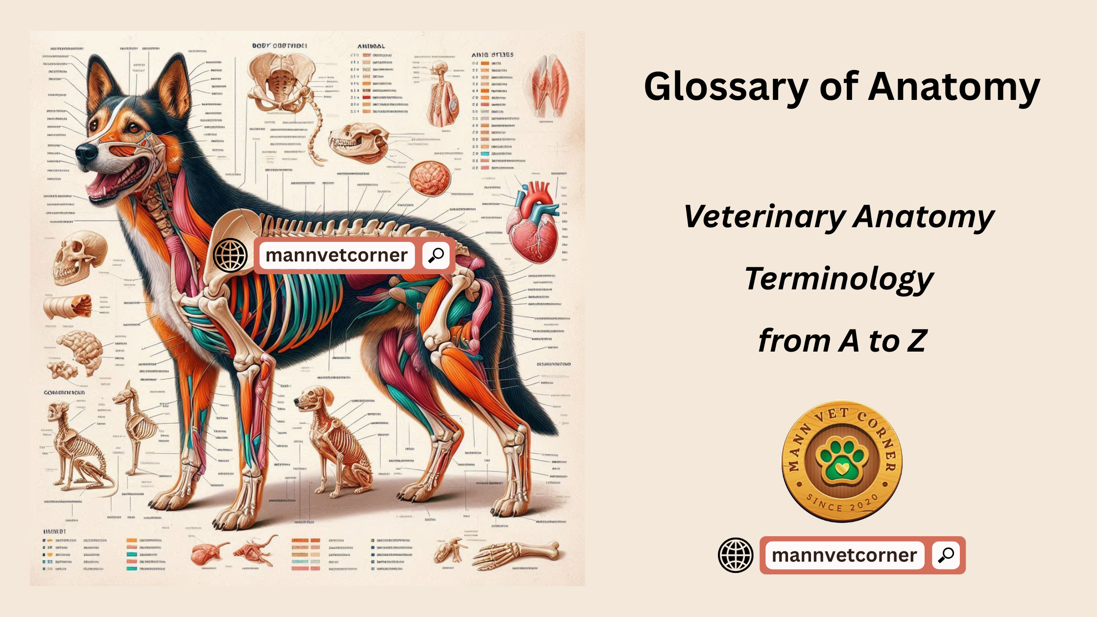
This glossary provides a foundational overview of veterinary anatomy terminology from A to Z, covering key terms across various anatomical systems and structures. While exhaustive detail for every species-specific term is beyond this scope, it includes essential vocabulary for understanding animal anatomy.
A
- Abdomen: The region of the body between the thorax and pelvis, housing digestive organs.
- Abomasum: The fourth stomach compartment in ruminants, functioning similarly to a non-ruminant stomach.
- Acetabulum: The pelvic socket where the femur articulates.
- Adipose tissue: Fat tissue that stores energy and provides insulation.
- Alveolus: A small cavity, such as the air sacs in the lungs or tooth sockets in the jaw.
- Anatomy: The scientific study of the structure of organisms.
- Anconeus muscle: A forearm muscle aiding elbow extension.
- Anus: The terminal opening of the digestive tract for waste expulsion.
- Aorta: The primary artery distributing oxygenated blood from the heart.
- Appendicular skeleton: The bones of the limbs and their girdles (e.g., shoulders and pelvis).
- Artery: A vessel carrying blood away from the heart.
- Articulation: A joint connecting two bones.
- Atlas: The first cervical vertebra supporting the skull.
- Atrium: One of the heart’s two upper chambers.
- Axial skeleton: The bones of the head, neck, and trunk.
B
- Biceps brachii: An upper arm muscle that flexes the elbow.
- Bile duct: A tube transporting bile from the liver to the small intestine.
- Bladder: An organ storing urine prior to excretion.
- Bone: Hard, dense connective tissue forming the skeleton.
- Brain: The central organ controlling bodily functions and processing sensory input.
- Bronchus: A tracheal branch leading to the lungs.
- Buccal cavity: The mouth cavity.
- Bursa: A fluid-filled sac reducing friction between moving body parts.
C
- Cannon bone: A lower leg bone in horses and other ungulates.
- Canine tooth: A pointed tooth for tearing food.
- Capillary: A minute blood vessel facilitating gas exchange.
- Cardiac muscle: Specialized muscle tissue of the heart.
- Carpus: The wrist joint in animals.
- Cartilage: Flexible connective tissue in joints and other structures.
- Cecum: A pouch at the large intestine’s start.
- Cerebellum: A brain region controlling balance and coordination.
- Cerebrum: The brain’s largest part, managing higher functions like thought and sensation.
- Cervical vertebrae: The neck’s spinal bones.
- Clavicle: The collarbone.
- Cochlea: A spiral inner ear structure for hearing.
- Colon: The large intestine.
- Cornea: The eye’s transparent front layer.
- Cranium: The skull portion enclosing the brain.
- Croup: The animal’s rump or buttocks.
D
- Dermis: The skin’s inner layer.
- Diaphragm: A muscle separating the chest and abdominal cavities, aiding respiration.
- Digestive system: Organs involved in food breakdown and nutrient absorption.
- Distal: Farther from the body’s center or attachment point.
- Dorsal: Relating to the back or upper body surface.
- Duodenum: The small intestine’s first segment.
E
- Ear: The organ of hearing and balance.
- Elbow: The joint connecting the humerus to the radius and ulna.
- Endocrine system: Glands producing hormones for bodily regulation.
- Epidermis: The skin’s outer layer.
- Epiglottis: A cartilage flap preventing food from entering the trachea during swallowing.
- Esophagus: The tube conveying food from mouth to stomach.
- Eustachian tube: A passage linking the middle ear to the throat.
F
- Facet: A smooth bone surface for articulation with another bone.
- Fascia: Connective tissue covering muscles and organs.
- Femur: The thigh bone.
- Fibula: The smaller lower leg bone.
- Follicle: A small sac, such as a hair follicle.
- Forelimb: An animal’s front leg.
- Frontal bone: A skull bone forming the forehead.
G
- Gallbladder: An organ storing bile.
- Gastric: Relating to the stomach.
- Gastrocnemius muscle: A calf muscle flexing the ankle.
- Genital system: The reproductive organs.
- Gingiva: The gums.
- Gland: An organ secreting substances like hormones or enzymes.
- Gluteal muscles: Buttock muscles.
H
- Hair: A filamentous skin growth.
- Heart: The organ pumping blood throughout the body.
- Hematopoiesis: Blood cell production, primarily in bone marrow.
- Humerus: The upper arm bone.
- Hypothalamus: A brain region regulating temperature, hunger, and thirst.
- Hyoid bone: A U-shaped neck bone supporting the tongue.
I
- Ileum: The small intestine’s final section.
- Incisor: A front tooth for cutting food.
- Inferior: Below another structure.
- Inguinal: Pertaining to the groin.
- Integumentary system: The skin and its appendages (e.g., hair, nails).
- Intercostal muscles: Muscles between ribs aiding respiration.
- Intestine: The digestive tract segment between stomach and anus.
J
- Jejunum: The small intestine’s middle portion.
- Joint: The connection point between two bones.
K
- Kidney: An organ filtering blood to produce urine.
- Knee: The joint between the femur and tibia.
L
- Lacrimal gland: A gland producing tears.
- Larynx: The voice box.
- Lateral: Away from the body’s midline.
- Ligament: Tissue connecting bones.
- Liver: An organ producing bile and detoxifying blood.
- Lumbar vertebrae: Lower back spinal bones.
- Lung: An organ enabling gas exchange.
M
- Mandible: The lower jawbone.
- Marrow: Soft bone tissue producing blood cells.
- Maxilla: The upper jawbone.
- Medial: Toward the body’s midline.
- Mediastinum: The space between the lungs.
- Meninges: Membranes covering the brain and spinal cord.
- Metacarpus: Hand or forefoot bones.
- Metatarsus: Foot or hindfoot bones.
- Molar: A back tooth for grinding.
- Muscle: Tissue contracting to produce movement.
N
- Naris: A nostril.
- Nasal cavity: The nose’s internal space.
- Neck: The region between head and trunk.
- Nerve: Fibers transmitting impulses.
- Nervous system: The brain, spinal cord, and nerves.
- Neuron: A nerve cell.
- Nipple: A mammary gland projection secreting milk.
O
- Occipital bone: A skull bone at the head’s back.
- Olfactory: Relating to smell.
- Omasum: The third stomach compartment in ruminants.
- Optic nerve: A nerve transmitting visual data from eye to brain.
- Orbit: The eye socket.
- Organ: A tissue group performing a specific function.
- Ovary: A female organ producing eggs.
P
- Palate: The mouth’s roof.
- Pancreas: An organ producing digestive enzymes and hormones.
- Patella: The kneecap.
- Pectoral: Relating to the chest.
- Pelvis: The hip bone structure.
- Penis: The male copulatory organ.
- Pericardium: The heart’s surrounding membrane.
- Peritoneum: The abdominal cavity’s lining membrane.
- Phalanges: Finger or toe bones.
- Pharynx: The throat.
- Pituitary gland: A hormone-regulating gland.
- Placenta: An organ linking fetus to uterus.
- Plasma: Blood’s liquid component.
- Pleura: The membrane lining the chest cavity and lungs.
- Pons: A brain region connecting cerebrum and cerebellum.
- Prostate gland: A male gland producing seminal fluid.
- Proximal: Nearer to the body’s center or attachment point.
Q
- Quadriceps femoris: Thigh muscles extending the knee.
R
- Radius: The forearm’s shorter bone.
- Rectum: The large intestine’s final section.
- Renal: Relating to the kidneys.
- Reproductive system: Organs for producing offspring.
- Respiratory system: Organs for breathing.
- Reticulum: The second stomach compartment in ruminants.
- Rib: A chest cage bone.
- Rumen: The first stomach compartment in ruminants.
S
- Sacrum: A triangular bone at the spine’s base.
- Salivary gland: A gland producing saliva.
- Scapula: The shoulder blade.
- Sebaceous gland: A gland secreting oil for skin lubrication.
- Semen: Fluid carrying sperm.
- Sensory organ: An organ detecting stimuli.
- Skeleton: The body’s bony framework.
- Skin: The body’s outer covering.
- Skull: The brain-enclosing bony structure.
- Small intestine: The digestive tract’s primary digestion and absorption site.
- Smooth muscle: Muscle in internal organ walls.
- Spinal cord: The nervous system’s vertebral column component.
- Spleen: An organ filtering blood and producing lymphocytes.
- Sternum: The breastbone.
- Stomach: An organ digesting food.
- Subcutaneous: Beneath the skin.
- Superior: Above another structure.
- Suture: A skull bone joint.
- Synovial joint: A freely movable joint.
T
- Tarsus: The ankle joint.
- Tendon: Tissue connecting muscle to bone.
- Testis: A male organ producing sperm.
- Thorax: The chest cavity.
- Thymus: A gland producing immune T-cells.
- Thyroid gland: A gland regulating metabolism via hormones.
- Tibia: The larger lower leg bone.
- Tongue: A muscular organ for taste and swallowing.
- Trachea: The windpipe.
- Triceps brachii: An upper arm muscle extending the elbow.
- Tympanic membrane: The eardrum.
U
- Ulna: The forearm’s longer bone.
- Umbilicus: The navel.
- Ureter: A tube from kidney to bladder.
- Urethra: A tube from bladder to exterior.
- Uterus: The womb for fetal development.
V
- Vagina: The female copulatory organ.
- Valve: A structure preventing fluid backflow.
- Vein: A vessel carrying blood to the heart.
- Vena cava: A major vein to the heart.
- Ventricle: One of the heart’s lower chambers.
- Vertebra: A spinal bone.
- Vestibule: A chamber, such as in the ear or reproductive system.
- Villus: A small intestinal projection for absorption.
- Viscera: Internal organs.
W
- Wither: The highest back point in horses, between shoulder blades.
X
- Xiphoid process: The sternum’s lower part.
Y
- Yolk sac: A membrane nourishing the embryo.
Z
- Zygomatic arch: The cheekbone.