Introduction
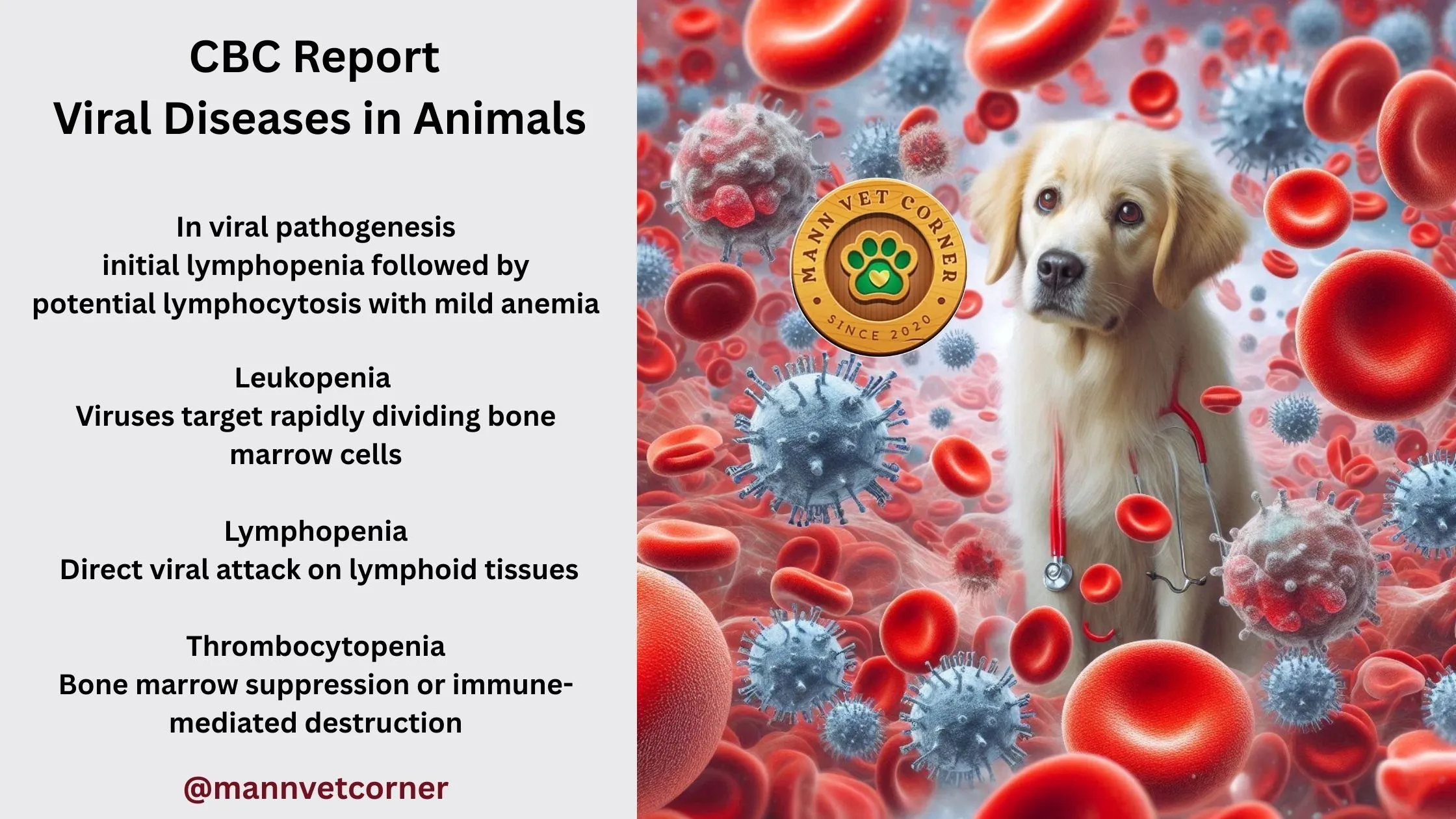
Viral infections in animals produce characteristic hematological changes that can be detected through comprehensive CBC analysis, serving as crucial diagnostic indicators for veterinary practitioners. During viral pathogenesis, the immune system responds with measurable alterations in white blood cell populations, including initial lymphopenia (decrease number of lymphocytes) followed by potential lymphocytosis (increase number of lymphocytes), while red blood cell parameters may show mild anemia (decrease number of RBCs) due to inflammatory processes and decreased erythropoiesis. Platelet counts often fluctuate, with thrombocytopenia (Low level of plateletes) commonly observed in acute viral infections such as parvovirus, distemper, and feline leukemia virus, reflecting both direct viral cytotoxicity and immune-mediated destruction.
The CBC differential count becomes particularly valuable in distinguishing viral from bacterial infections, as viral etiologies typically present with relative lymphocytosis and absence of left shift neutrophilia (means there is a higher than expected percentage of immature neutrophils from bone marrow), though exceptions exist in severe systemic viral diseases where secondary bacterial complications may develop. Additionally, certain viral infections like feline immunodeficiency virus and bovine leukemia virus can cause persistent hematological abnormalities including progressive lymphopenia or abnormal lymphocyte morphology, making serial CBC monitoring essential for disease progression assessment and treatment response evaluation in affected animals.
Viral Attacks: When Lymphocytes Crash
Lymphocytes are the immune system’s strategists, orchestrating adaptive immunity via T-cells, B-cells, and natural killer cells. Viruses, however, are cunning saboteurs, targeting lymphocytes to cripple immunity. A sharp drop in lymphocyte counts (lymphopenia) is a hallmark of viral infections, often detectable before clinical signs escalate.
Cattle: BVDV’s Devastating Trick
Bovine Viral Diarrhea Virus (BVDV) is a notorious bovine pathogen that decimates lymphocyte populations, causing mucosal ulcers, abortion in pregnant cows, and persistently infected (PI) calves if the fetus is exposed in utero. Lymphopenia (<2,000/μL) is a consistent finding, reflecting viral destruction of lymphoid tissues. PI calves, born immunotolerant, shed BVDV lifelong, posing a herd-wide threat.
Case Study: BVDV Outbreak
A beef cattle herd experienced abortions and diarrhea in calves. Bloodwork from affected cows showed lymphopenia (1,500/μL). PCR confirmed BVDV, and testing identified two PI calves, which were culled to halt transmission. Lymphopenia guided the diagnostic workup.
Cats: FPV’s Deadly Strike
Feline Panleukopenia Virus (FPV), a parvovirus, devastates cats by obliterating WBCs, earning its name “panleukopenia” (low all WBCs). Counts can plummet to <1,000 cells/μL, with lymphopenia and neutropenia reflecting bone marrow suppression. FPV also destroys intestinal crypt cells, causing bloody diarrhea and dehydration. Kittens are especially vulnerable, with mortality rates approaching 90% without intensive care.
Case Study: Feline Panleukopenia
A 12-week-old stray kitten presented with vomiting, diarrhea, and fever. WBC count was 800/μL, with 10% lymphocytes and 20% neutrophils. FPV antigen testing was positive, and aggressive fluid therapy saved the kitten. The panleukopenia was the diagnostic linchpin.
Chickens: Marek’s Viral Treason
Marek’s Disease, caused by a herpesvirus, is a poultry nightmare, inducing tumors and immune suppression. WBC patterns vary: some birds develop lymphocytosis from cancerous lymphocytes, while others show lymphopenia due to lymphoid depletion. Feather follicle tumors and paralysis are late signs, making early WBC analysis critical.
Did You Know? In dogs with canine distemper, atypical lymphocytes—large, reactive cells with irregular nuclei—appear before neurological signs like seizures, offering an early diagnostic clue.
Major CBC Patterns in Viral Diseases:
1. Classic Viral Pattern: Leukopenia + Lymphopenia + Thrombocytopenia
- This triad is seen in diseases like Parvovirus, BVD, and MERS-CoV
- Indicates bone marrow suppression and immune system targeting
2. Species-Specific Viral Signatures:
- Dogs: Parvovirus shows severe leukopenia (<2,000/μL) – almost pathognomonic
- Cats: Retroviral infections (FeLV/FIV) cause persistent progressive CBC decline
- Cattle: BVD shows the classic triad with immunosuppression
- Horses: EIA causes anemia + thrombocytopenia pattern
- Camels: Respiratory viruses typically show leukopenia + lymphopenia
3. Diagnostic Reasoning Behind CBC Changes:
- Leukopenia: Viruses target rapidly dividing bone marrow cells
- Lymphopenia: Direct viral attack on lymphoid tissues
- Thrombocytopenia: Bone marrow suppression or immune-mediated destruction
- Anemia: Usually secondary from chronic disease or hemolysis
4. Key Diagnostic Differentiators:
- Viral vs Bacterial: Viral diseases typically show leukopenia without left shift, while bacterial infections show neutrophilia with left shift
- Acute vs Chronic: Acute viral infections show rapid CBC decline, chronic infections show persistent mild abnormalities
5. Prognostic Value:
- Severe leukopenia (<2,000/μL) indicates poor prognosis
- Recovery of WBC counts within 7-10 days suggests good prognosis
- Secondary bacterial infections (shown by neutrophilia) complicate viral diseases
The table demonstrates that CBC analysis is crucial for early viral disease detection, especially when combined with clinical signs and confirmatory testing like PCR, serology, or antigen tests.
Comprehensive Table of Viral Diseases with CBC Diagnostic Findings
| Disease | Host | Clinical Findings | CBC Findings | Reasons for CBC Changes | Diagnosis |
|---|---|---|---|---|---|
| Canine Parvovirus | Dogs | Bloody diarrhea, vomiting, dehydration, lethargy | Severe leukopenia (WBC: 1,500/μL), neutropenia (<500/μL), lymphopenia | Virus destroys rapidly dividing cells in bone marrow and intestines | Clinical signs + CBC pattern + fecal antigen test |
| Feline Leukemia Virus (FeLV) | Cats | Weight loss, recurrent infections, lymphadenopathy | Progressive anemia (PCV: 18%), leukopenia (3,000/μL), thrombocytopenia | Virus causes bone marrow suppression and immunosuppression | SNAP test + persistent CBC abnormalities |
| Feline Immunodeficiency Virus (FIV) | Cats | Recurrent abscesses, dental disease, chronic infections | Neutropenia (1,500/μL), lymphopenia, mild anemia | Virus attacks CD4+ T cells causing immunosuppression | SNAP test + chronic neutropenia pattern |
| Bovine Viral Diarrhea (BVD) | Cattle | Diarrhea in calves, respiratory signs, immunosuppression | Leukopenia (2,500/μL), lymphopenia, thrombocytopenia (100,000/μL) | Virus causes bone marrow suppression and platelet destruction | Antigen testing + characteristic CBC pattern |
| Equine Infectious Anemia (EIA) | Horses | Fever, weight loss, dependent edema, weakness | Anemia (PCV: 20%), thrombocytopenia (80,000/μL), lymphopenia | Virus causes immune-mediated destruction of RBCs and platelets | Coggins test (ELISA) + CBC findings |
| Equine Herpesvirus-1 (EHV-1) | Horses | Respiratory disease, neurologic signs in mares | Leukopenia (3,500/μL), lymphopenia, thrombocytopenia | Virus causes lymphoid tissue destruction and platelet consumption | PCR + serology + CBC pattern |
| MERS-CoV | Camels | Respiratory distress, nasal discharge, fever | Leukopenia (3,500/μL), lymphopenia, mild thrombocytopenia | Virus targets immune cells and causes systemic inflammation | RT-PCR + clinical signs + CBC |
| Camel Pox | Camels | Skin lesions, fever, respiratory signs | Initial leukocytosis (16,000/μL), then leukopenia, lymphopenia | Early inflammatory response followed by viral immunosuppression | Viral isolation + CBC progression |
| Bovine Respiratory Syncytial Virus (BRSV) | Cattle | Coughing, respiratory distress in young calves | Leukopenia (4,000/μL), lymphopenia, normal RBC initially | Virus causes respiratory tract inflammation and immune suppression | Serology + CBC findings + clinical signs |
| Bovine Leukosis (Enzootic) | Cattle | Enlarged lymph nodes, weight loss, tumor formation | Lymphocytosis (>10,000/μL), atypical lymphocytes, mild anemia | Virus causes malignant transformation of B lymphocytes | Serology + persistent lymphocytosis |
| West Nile Virus | Horses | Ataxia, weakness, muscle fasciculations, neurologic signs | Normal to slightly elevated WBC, mild lymphocytosis | Virus primarily affects nervous system with minimal blood changes | Serology + clinical neurologic signs |
| Bluetongue Disease | Camels, Sheep | Oral ulcers, lameness, coronitis, tongue swelling | Leukopenia (4,000/μL), thrombocytopenia, mild anemia | Virus causes vasculitis and bone marrow suppression | RT-PCR + characteristic CBC + clinical signs |
| Rift Valley Fever | Camels, Ruminants | Fever, weakness, nasal discharge, hemorrhages | Leukopenia, thrombocytopenia, mild anemia | Virus causes widespread tissue necrosis and coagulopathy | Serology + CBC pattern + clinical signs |
| Foot and Mouth Disease | Camels, Ruminants | Vesicular lesions on feet and mouth, fever | Leukopenia initially, then recovery to normal | Initial viral suppression followed by recovery response | ELISA + virus isolation + clinical lesions |
| Ovine Progressive Pneumonia (OPP) | Goats | Progressive respiratory difficulty, weight loss | Normal to slightly elevated WBC, chronic disease anemia | Chronic viral infection causes persistent low-grade inflammation | Serology + chronic CBC changes |
Key CBC Changes in Viral Diseases
Primary Patterns:
1. Leukopenia (Most Common)
- Mechanism: Viruses often target bone marrow stem cells and lymphoid tissues
- Significance: Indicates viral suppression of immune cell production
- Examples: Parvovirus, FeLV, BVD, MERS-CoV
2. Lymphopenia
- Mechanism: Many viruses specifically target lymphocytes or lymphoid organs
- Significance: Reflects immunosuppression and viral tropism for immune cells
- Examples: FIV, EHV-1, BRSV, Bluetongue
3. Thrombocytopenia
- Mechanism: Viral-induced bone marrow suppression or immune-mediated platelet destruction
- Significance: Indicates systemic viral effects on hematopoiesis
- Examples: EIA, BVD, Rift Valley Fever
4. Anemia (Secondary)
- Mechanism: Chronic disease, immune-mediated hemolysis, or bone marrow suppression
- Significance: Indicates chronic viral infection or immune-mediated destruction
- Examples: FeLV, EIA, chronic viral infections
Diagnostic Significance:
Acute Viral Infections:
- Rapid onset leukopenia with lymphopenia
- Often accompanied by thrombocytopenia
- May show recovery pattern if animal survives
Chronic Viral Infections:
- Persistent mild to moderate CBC abnormalities
- Progressive anemia in immunosuppressive viruses
- Secondary bacterial infections may cause neutrophilia
Immunosuppressive Viruses:
- Profound and persistent leukopenia
- Increased susceptibility to secondary infections
- Poor prognosis indicators
Species-Specific Considerations:
Dogs:
- Parvovirus: Severe leukopenia is pathognomonic
- Stress leukogram may mask viral effects in some cases
Cats:
- Retroviral infections (FeLV/FIV): Persistent CBC abnormalities
- Secondary infections common due to immunosuppression
Cattle:
- BVD: Classic triad of leukopenia, lymphopenia, thrombocytopenia
- Immunosuppression leads to secondary bacterial infections
Horses:
- EIA: Anemia with thrombocytopenia is characteristic
- Neurotropic viruses may have minimal CBC changes
Camels:
- Respiratory viruses: Leukopenia with lymphopenia pattern
- Systemic viruses: More pronounced CBC abnormalities
Diagnostic Approach:
- Pattern Recognition: Identify characteristic viral CBC patterns
- Clinical Correlation: Match CBC findings with clinical signs
- Confirmatory Testing: Use specific viral tests (serology, PCR, antigen)
- Monitoring: Track CBC changes over time for prognosis
Key Diagnostic Clues:
Highly Suggestive of Viral Disease:
- Leukopenia + Lymphopenia + Clinical signs
- Thrombocytopenia in young animals
- Lack of left shift despite illness
- Progressive CBC deterioration
Differentiating from Bacterial Disease:
- Bacterial: Usually neutrophilia with left shift
- Viral: Usually leukopenia without left shift
- Mixed infections: May show both patterns
Prognostic Indicators:
Poor Prognosis:
- Severe persistent leukopenia (<2,000/μL)
- Progressive thrombocytopenia
- Development of secondary bacterial infections
Good Prognosis:
- Mild CBC changes with clinical improvement
- Recovery of WBC counts within 7-10 days
- Maintenance of platelet counts
Summary
Viral diseases in animals typically cause leukopenia, lymphopenia, and thrombocytopenia due to:
- Bone marrow suppression
- Direct viral cytotoxicity to immune cells
- Immune-mediated destruction of blood cells
- Systemic inflammatory responses
The CBC serves as a crucial diagnostic tool when combined with clinical signs and confirmatory viral testing. The pattern of leukopenia with lymphopenia is the hallmark of most viral infections, distinguishing them from bacterial diseases which typically cause neutrophilia. Early recognition of these patterns allows for prompt diagnosis, appropriate supportive care, and prevention of secondary complications.

