

Quick Answer:
What does a CBC tell you in animal disease diagnosis?
A complete blood count (CBC) measures red cells, white cells, and platelets. In animal disease diagnosis the pattern matters more than any single number: low white cells with low neutrophils and lymphocytes suggest viral diseases such as parvovirus, panleukopenia, distemper, and PPR; a high white count with neutrophilia points to bacterial or inflammatory disease such as strangles and FIP; and severe anemia with thrombocytopenia in a tick-exposed bovine suggests babesiosis. A CBC supports a diagnosis and guides urgency, but confirmatory tests such as PCR, antigen tests, and blood smears make it definitive.
Key Takeaways
- Read patterns, not single values. Leukopenia + neutropenia + lymphopenia is the classic viral pattern; neutrophilia + lymphopenia is the classic inflammatory pattern.
- Use species-specific reference intervals. A value that is normal in a cow can be abnormal in a cat, and kittens, puppies and lambs differ from adults.
- Dehydration hides anemia. A hematocrit of 32% in a vomiting kitten may be lower after rehydration.
- Always review the blood smear. Toxic neutrophils, left shift, atypical lymphocytes and blood parasites are found by the microscope, not the analyzer.
- CBC is a screening and monitoring tool. Confirm with PCR, antigen ELISA, culture, serology or smear examination.
Introduction: Why the CBC Is the First Test Veterinarians Reach For
When a puppy passes bloody diarrhea, a foal develops swollen jaw lymph nodes, or a cow passes red urine in tick season, the clinician needs answers fast. A complete blood count (CBC)is one of the quickest, most affordable, and most informative tests available in veterinary practice. Within minutes it shows whether the animal is fighting an infection, losing red cells, running out of white cells, or bleeding platelets away.
Yet a CBC report is only as useful as the person reading it. A number such as “WBC 1.2 × 10³/µL” means nothing until it is compared with the correct species reference interval, the animal’s age and hydration status, and the clinical signs in front of you. The real skill in animal disease diagnosis is pattern recognition: seeing how white cells, red cells and platelets move together and matching that fingerprint to the diseases that produce it.
This guide walks through seven important animal diseases and the CBC fingerprint each one leaves: feline panleukopenia, canine parvovirus, canine distemper, feline infectious peritonitis (FIP), equine strangles, peste des petits ruminants (PPR) and bovine babesiosis. Each section pairs typical clinical signs with a representative CBC report, explains why the numbers change, and lists the confirmatory tests that should follow. Whether you are a veterinary student, a practicing veterinarian, a livestock farmer working with your vet, or a pet owner trying to understand a lab report, you will leave able to read a CBC with more confidence.
What Is a CBC in Veterinary Medicine?
A complete blood count analyzes the cellular components of blood collected in an EDTA (purple-top) tube. It is normally reported in three groups: the red cell line, the white cell line (with a differential), and platelets. Modern hematology analyzers report most values automatically, but a trained person should confirm them with a stained blood smear, particularly in cats, horses and ruminants, where automated counts can be misleading.
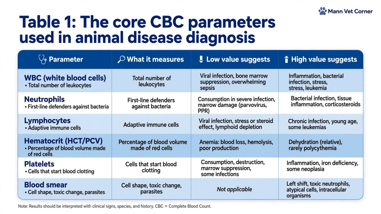
The core CBC parameters and what they mean
Table 1: The core CBC parameters used in animal disease diagnosis.

Reference intervals differ between species
One of the most common interpretation errors is applying a dog range to a cat or a horse. Species differ, and so do age groups and even breeds (for example, sighthounds normally have a higher hematocrit and a lower white count).
Table 2: Approximate adult reference values of common species. 
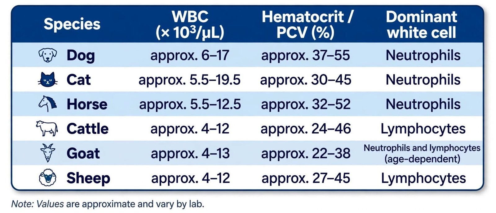
Notice that in healthy cattle and sheep, lymphocytes usually outnumber neutrophils. A cow with a “normal” neutrophil percentage may therefore already have a relative neutrophilia. This is why livestock CBCs should never be read with small-animal rules of thumb.
A Step-by-Step Method for Reading a CBC Report
Experienced clinicians read a CBC the same way every time. A fixed routine prevents anchoring on one dramatic value and missing the rest of the story.
- Confirm the patient and the sample. Check species, age, sample tube, clotting or clumping flags and time since collection.
- Look at the red cell line first. Assess hematocrit, then ask whether hydration status is distorting it. Check for regeneration signs on the smear.
- Read the total WBC. Is it low (leukopenia), normal, or high (leukocytosis) for that species and age?
- Break it down with the differential. Which cell line is responsible: neutrophils, lymphocytes, monocytes or eosinophils?
- Check platelets and confirm on the smear. Automated counts can be falsely low when platelets clump, particularly in cats.
- Examine the blood smear. Look for left shift, toxic change, atypical lymphocytes, inclusion bodies and blood parasites.
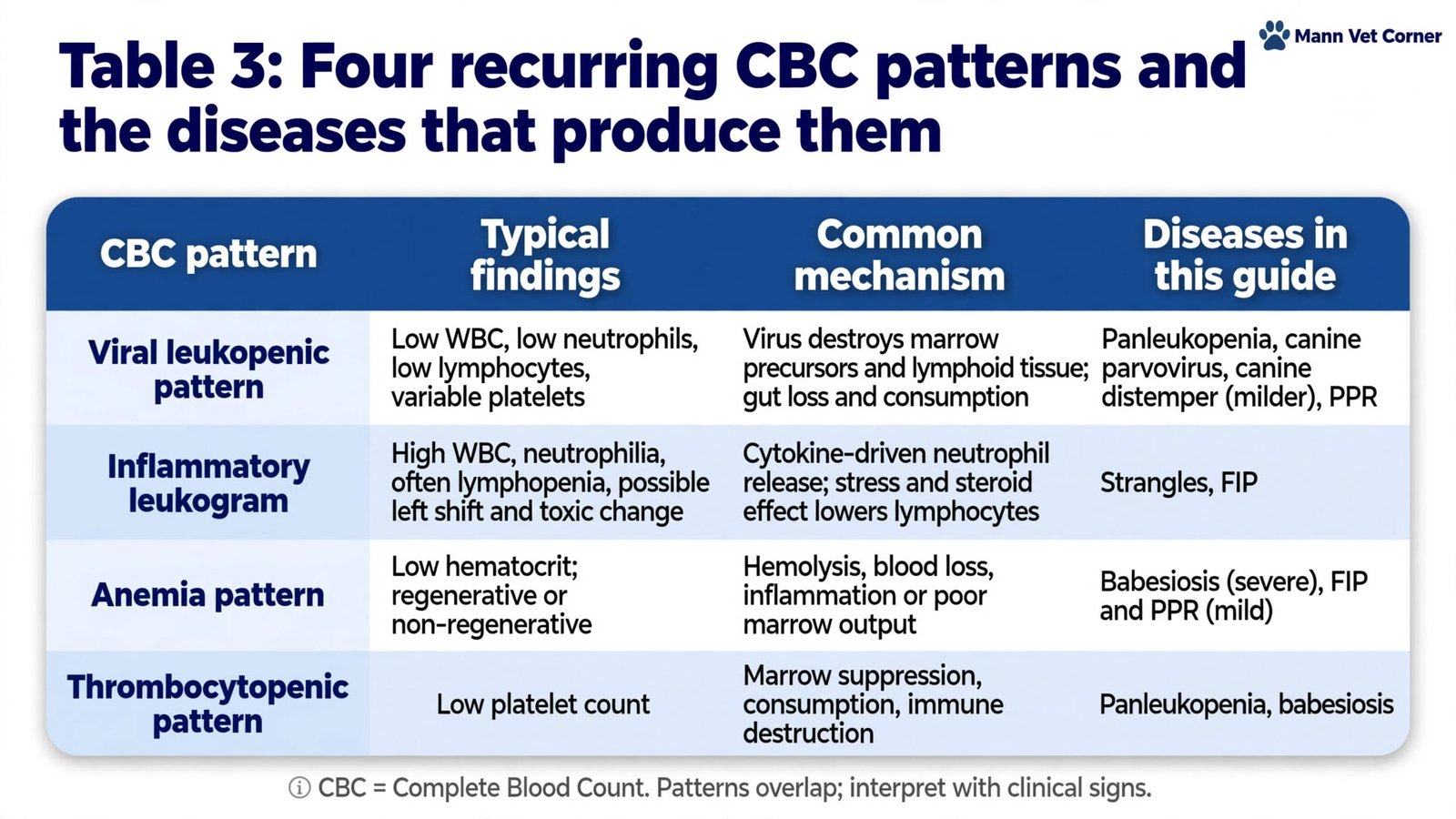
The Four Big CBC Patterns in Animal Disease
Before looking at individual diseases, it helps to know the four broad patterns that appear again and again in infectious disease work.
Table 3: Four recurring CBC patterns and the diseases that produce them.
CBC Findings in Seven Important Animal Diseases
Section A: Small Animal Diseases (Cats and Dogs)
1. Feline Panleukopenia (FPV) in Kittens
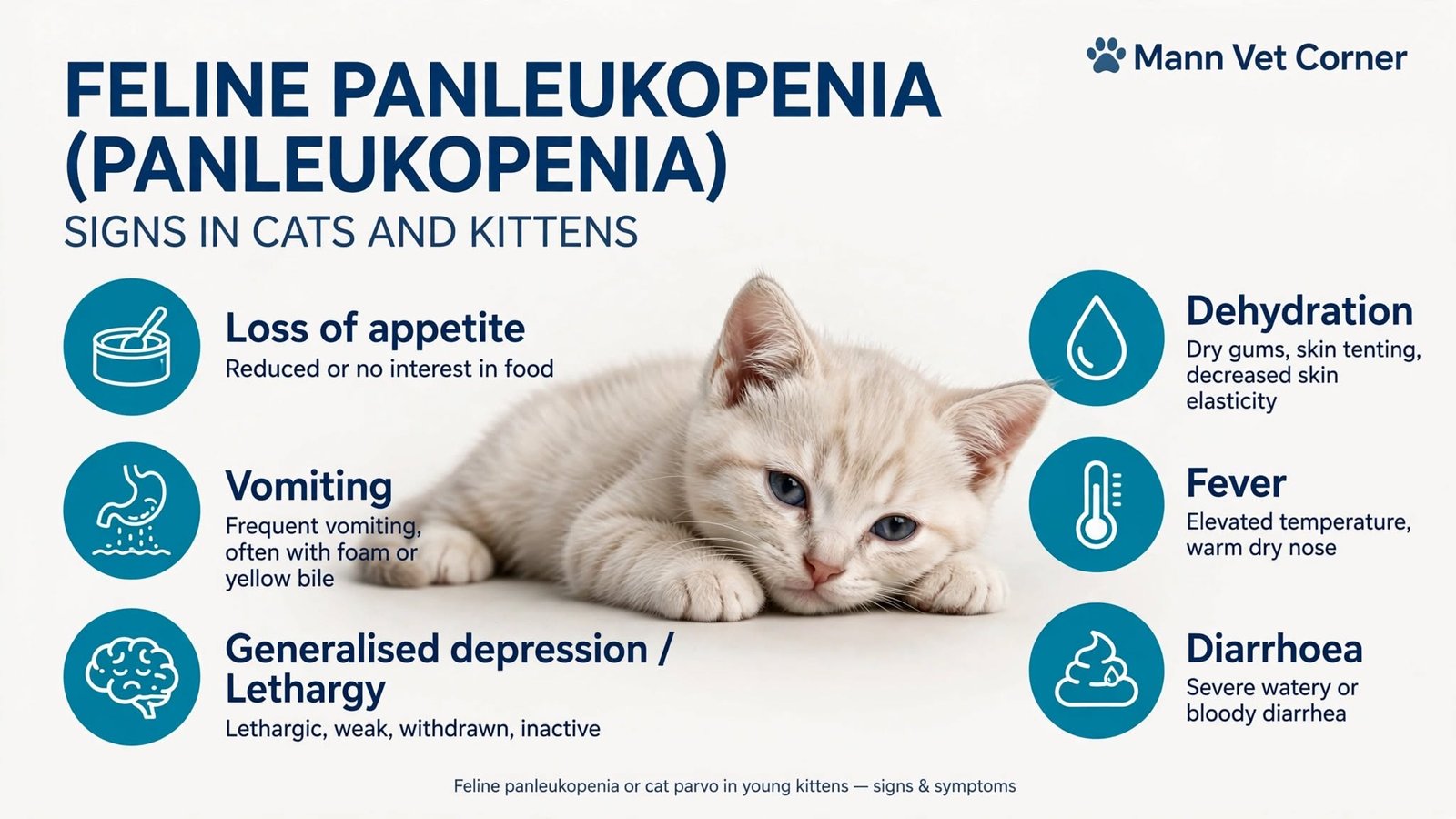
Clinical signs of feline panleukopenia
Feline panleukopenia, also called feline parvovirus or feline distemper, typically causes loss of appetite, vomiting (often foamy or bile-stained), fever, watery or bloody diarrhea, dehydration, and lethargy. In a young kitten, hypothermia usually signals advanced disease and circulatory compromise, so it is a red flag rather than a minor detail.
CBC results and interpretation
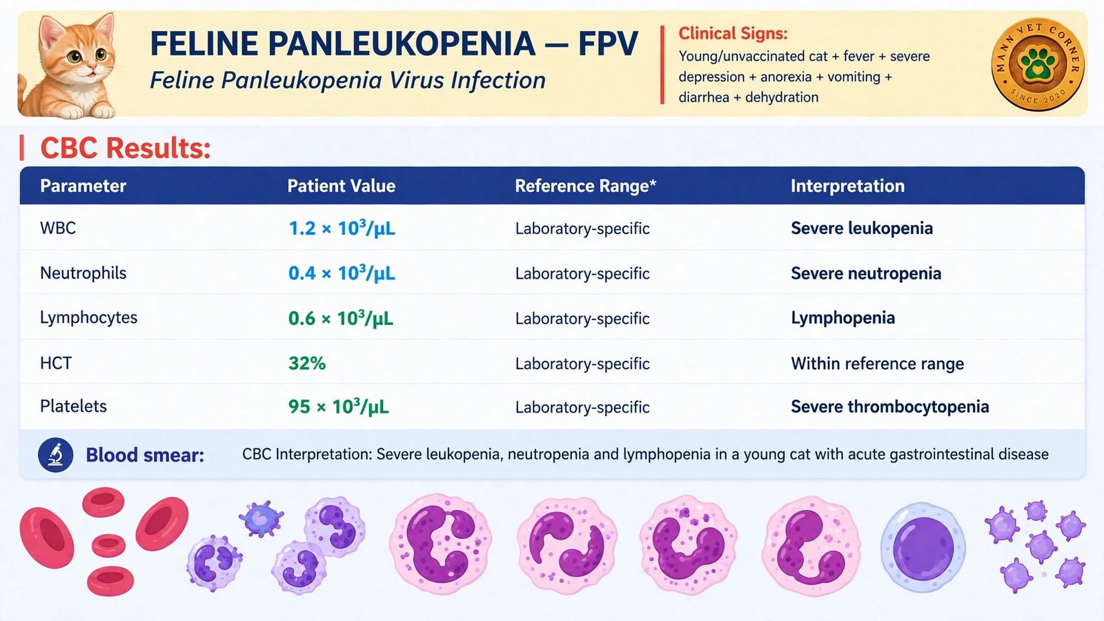
Table 4: Representative CBC in feline panleukopenia. Reference ranges are those printed on the case report.
Blood smear: Neutropenia may make a differential difficult to count; look for toxic change in surviving neutrophils and for platelet clumping before accepting a low platelet number.
Why the CBC looks this way
Feline parvovirus replicates in rapidly dividing cells: the intestinal crypt epithelium, bone marrow progenitors, and lymphoid tissue. Destruction of myeloid precursors causes profound neutropenia, lymphoid depletion causes lymphopenia, and hemorrhagic enteritis with consumption drives platelets down. Together they create the textbook viral leukopenic pattern that gives the disease its name.
The “normal” hematocrit of 32% is deceptive. Vomiting and diarrhea shrink plasma volume and concentrate red cells. Once the kitten is rehydrated, the hematocrit can fall, so it should be rechecked after fluid therapy rather than treated as reassuring.
2. Canine Parvovirus (CPV) Enteritis in Puppies
Clinical signs of canine parvovirus
Canine parvovirus causes loss of appetite, severe repeated vomiting, foul-smelling bloody diarrhea, abdominal pain, dehydration, fever, and lethargy. Puppies with incomplete vaccination are most at risk, and some breeds, such as Rottweilers, Dobermans, and pit bull types, are reported to be predisposed.
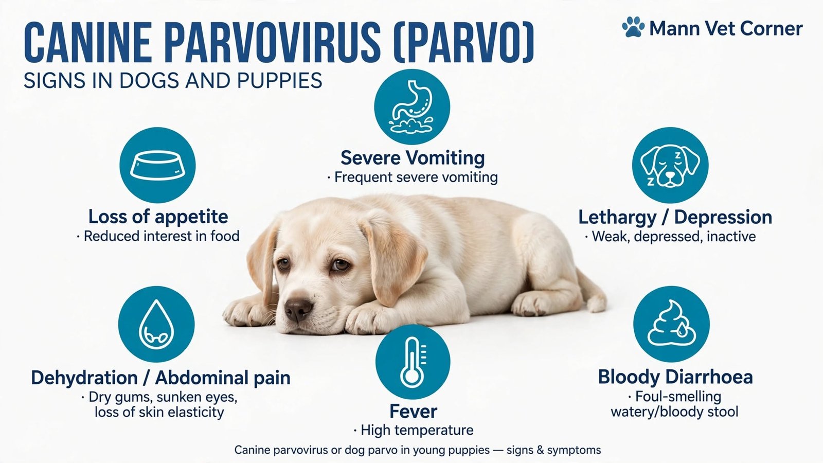
CBC results and interpretation
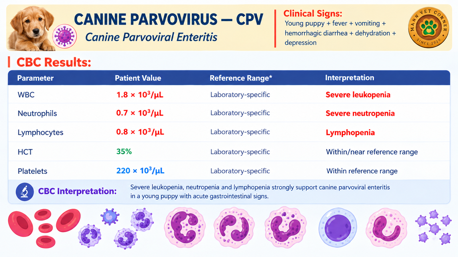
Table 5: Representative CBC in canine parvoviral enteritis.
Blood smear: Look for toxic changes and left shift in any remaining neutrophils. Their presence suggests bacterial translocation from the damaged intestine.
Why the CBC looks this way
Leukopenia, neutropenia and lymphopenia together strongly support canine parvoviral enteritis in a young puppy with acute gastrointestinal signs. The virus destroys intestinal crypt cells and the marrow and lymphoid cells that produce white blood cells, while damaged gut lining allows bacteria to enter the bloodstream and consume neutrophils.
Compare this puppy with the kitten above: the platelet count here is normal. That is a useful reminder that no single parameter is mandatory. The pattern of white cells, not the platelets, carries the diagnostic weight in this case.
3. Canine Distemper Virus (CDV) Infection
Case snapshot: A young, unvaccinated dog presents with fever, oculonasal discharge, cough, diarrhea and neurological signs.
Clinical signs of canine distemper
Canine distemper is a multisystem disease. Early signs include biphasic fever and purulent nasal and eye discharge, followed by coughing and breathing difficulty, vomiting and diarrhea. Later, seizures, twitching (myoclonus) and circling may develop, and hardening of the nose and foot pads (hard pad disease) can appear in some dogs.
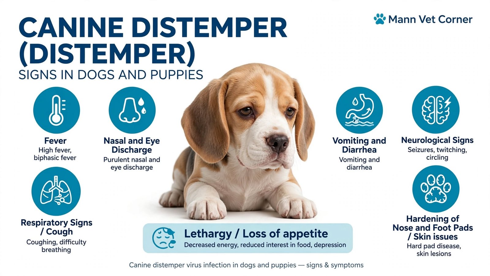
CBC results and interpretation
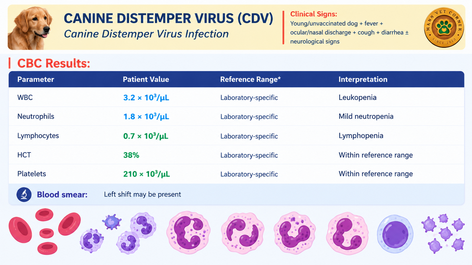
Table 6: Representative CBC in canine distemper virus infection.
Blood smear: A left shift may be present. In the early viremic phase, distemper inclusion bodies are occasionally seen in circulating red cells, neutrophils or lymphocytes, but their absence does not rule out infection.
Why the CBC looks this way
Canine distemper virus is lymphotropic. It infects lymphoid tissue early, causing lymphocyte destruction and profound immunosuppression, which is why lymphopenia is the most consistent CBC finding. The leukopenia is usually less dramatic than in parvovirus infection, because distemper does not destroy the intestinal and marrow compartments to the same degree.
Immunosuppression is also why secondary bacterial pneumonia and enteritis are so common in distemper. A dog whose CBC later swings to a neutrophilic leukocytosis may have developed such a secondary infection.
4. Feline Infectious Peritonitis (FIP)
Clinical signs of feline infectious peritonitis
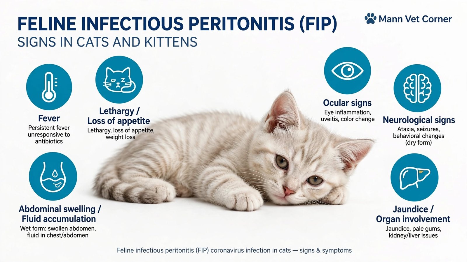
FIP, caused by a mutated feline coronavirus, appears as a “wet” (effusive) form with abdominal or chest fluid, or a “dry” (non-effusive) form with granulomatous lesions. Common signs include persistent fever, lethargy, weight loss, abdominal swelling, jaundice, ocular changes such as uveitis, and neurological signs such as ataxia or seizures.
CBC results and interpretation
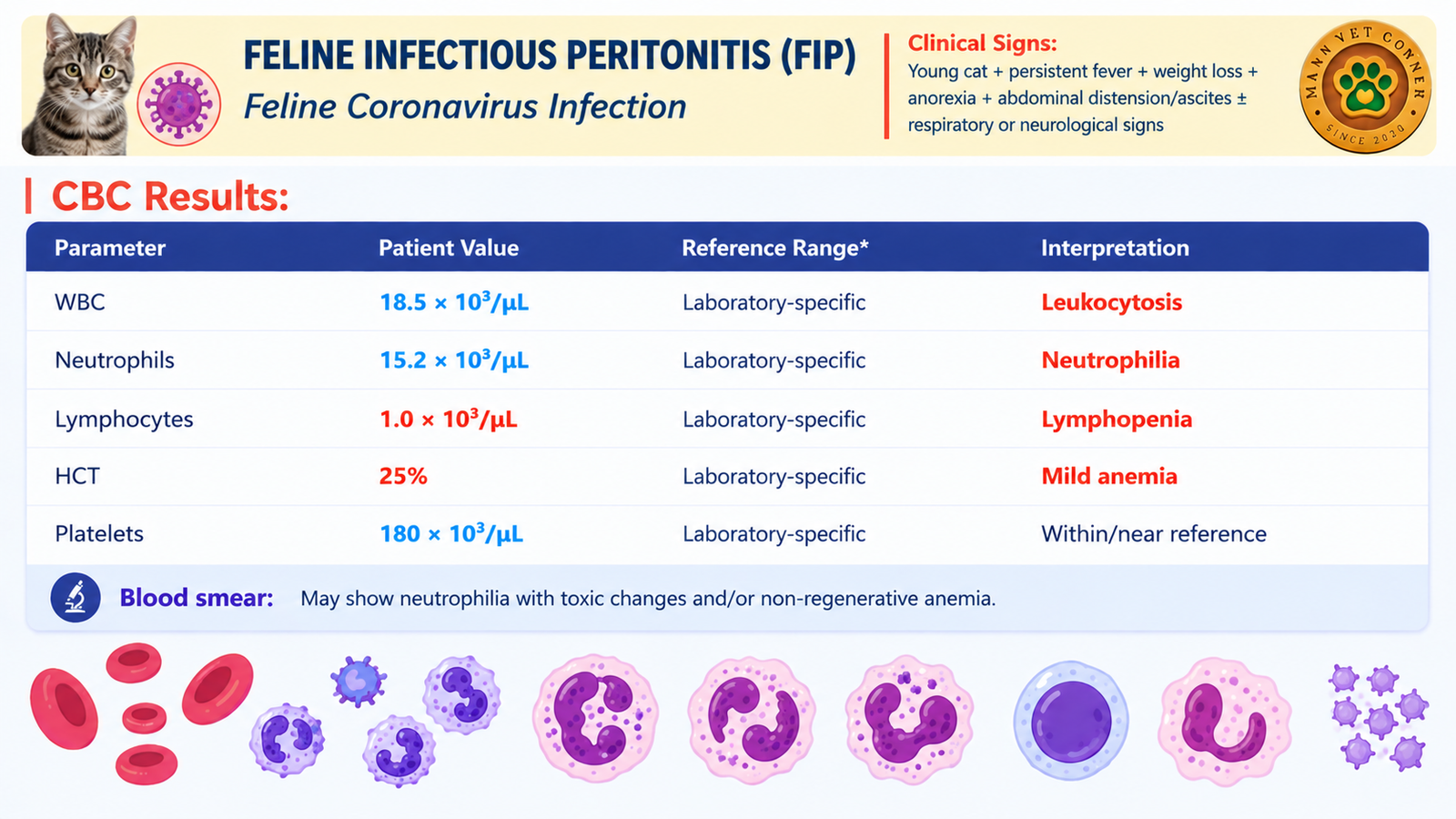
Table 7: Representative CBC in feline infectious peritonitis.
Blood smear: Neutrophils may show toxic changes, and the anemia is often non-regenerative (anemia of inflammatory disease).
Why the CBC looks this way
The FIP CBC shows an inflammatory leukogram: neutrophilia driven by cytokines, lymphopenia from stress and immune dysregulation, and a mild non-regenerative anemia caused by chronic inflammation. None of these findings is specific. Many bacterial infections, abscesses and neoplasms produce a similar picture, which is why FIP is diagnosed by combining the CBC with other evidence and never by the CBC alone.
Interestingly, the CBC here separates FIP from the parvoviral diseases above: the white count is high rather than low. The direction of the white cell count is the first branch point in the diagnostic tree.
Section B: Equine Disease
5. Equine Strangles (Streptococcus equi Infection)
Clinical signs of strangles
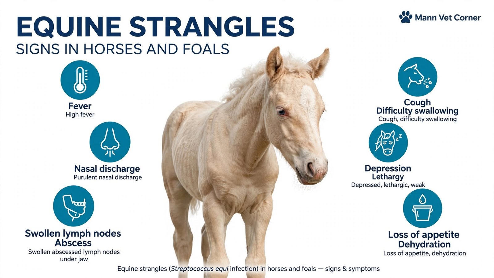
Strangles is a highly contagious upper respiratory infection caused by Streptococcus equi subspecies equi. Classic signs are high fever, thick purulent nasal discharge, painful swelling of the lymph nodes under the jaw and behind the throat, cough, dysphagia, depression and loss of appetite. The lymph node swelling may compress the airway, which gives the disease its name.
CBC results and interpretation
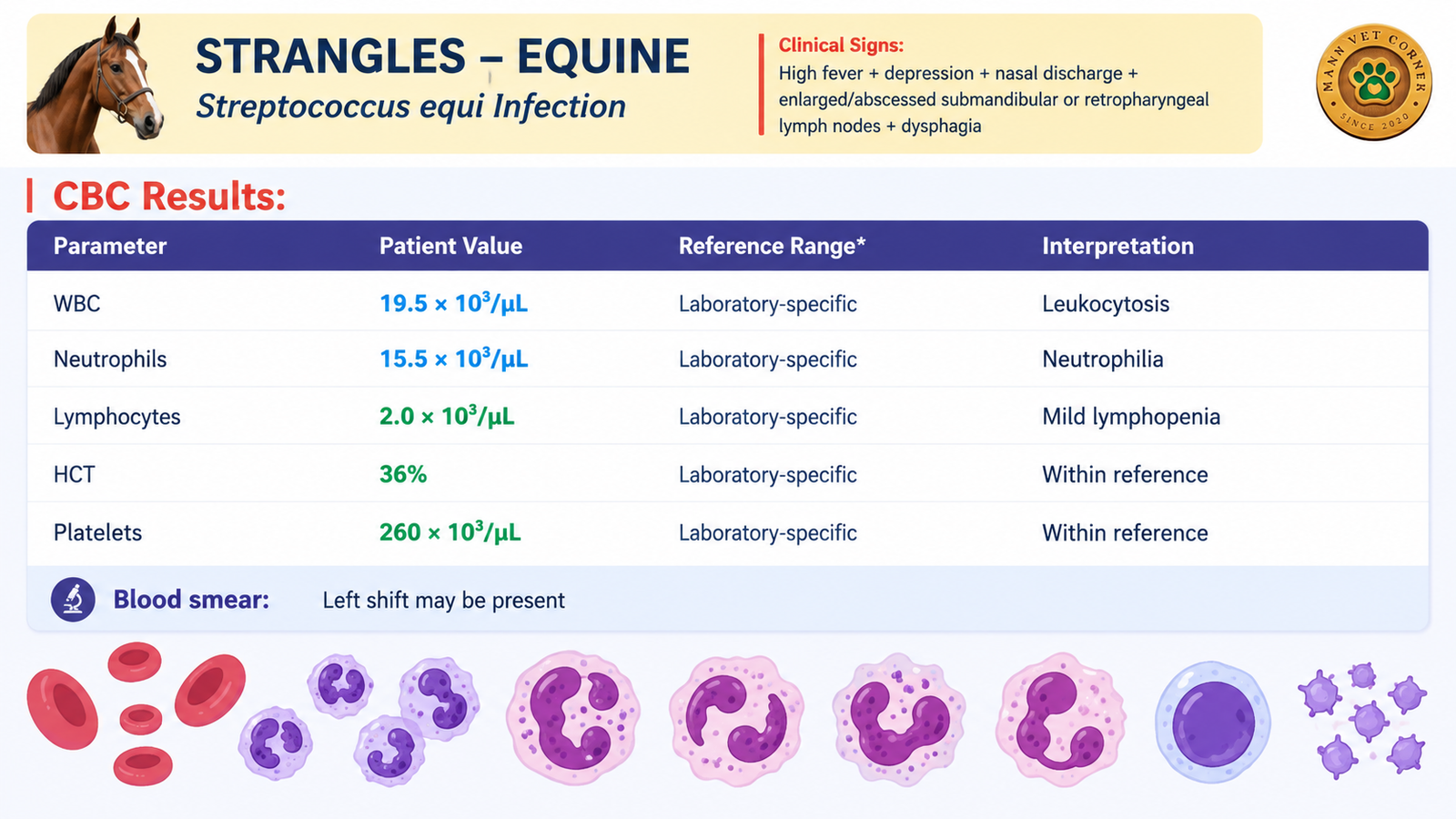
Table 8: Representative CBC in equine strangles. Whether 2.0 × 10³/µL lymphocytes counts as decreased.
Blood smear: A left shift and toxic neutrophils may be present, consistent with a bacterial infection.
Why the CBC looks this way
Strangles produces a classic bacterial inflammatory leukogram. The bone marrow releases large numbers of neutrophils in response to Streptococcus equi and abscess formation, so the total white count and the neutrophil count both rise. Lymphocytes are often normal to mildly reduced because of stress and inflammatory mediators.
The CBC is normally paired with acute-phase markers. Elevated plasma fibrinogen (often well above the upper reference limit) and elevated serum amyloid A support active inflammation, and both are useful for monitoring recovery. Anemia of inflammatory disease may develop in prolonged cases.
Section C: Small Ruminant Disease
6. Peste des Petits Ruminants (PPR) in Goats and Sheep
Clinical signs of PPR
PPR, caused by a morbillivirus, is a devastating disease of goats and sheep. Signs include high fever, watery to purulent nasal and eye discharge, mouth ulcers and stomatitis, cough and labored breathing, profuse watery diarrhea, dehydration and severe depression. Young animals and naive flocks suffer the highest mortality.
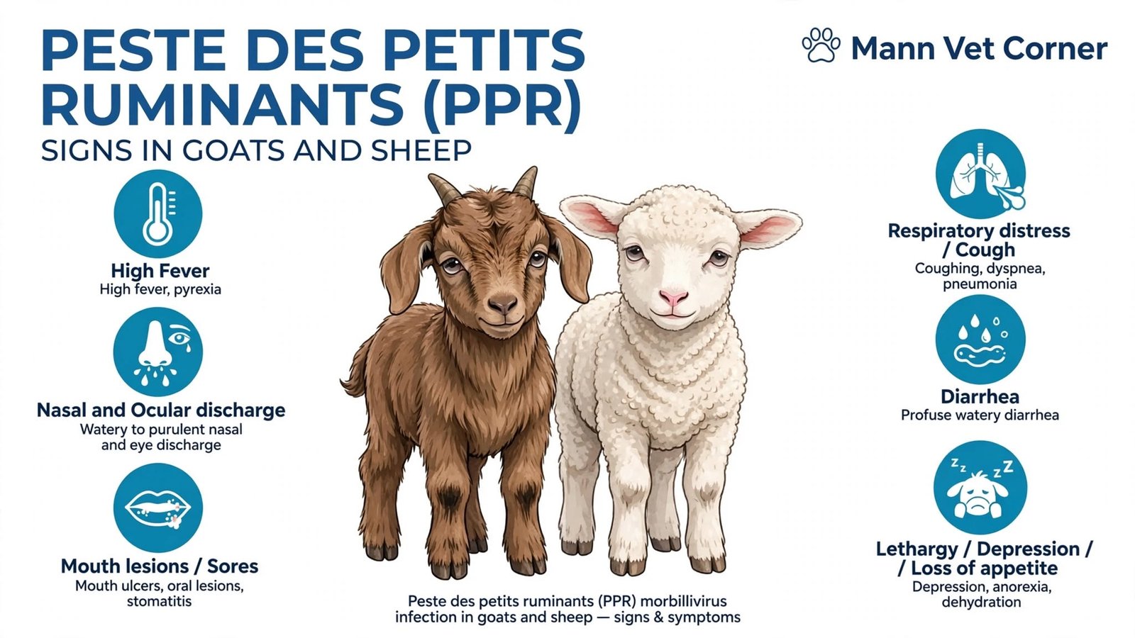
CBC results and interpretation
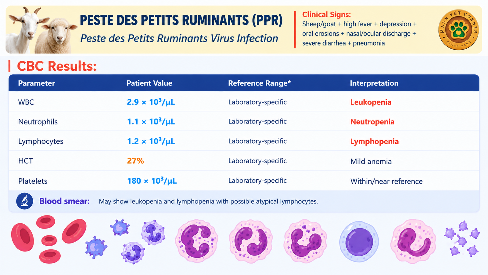
Table 9: Representative CBC in PPR. A hematocrit of 27% is at the low end.
Blood smear: Leukopenia and lymphopenia are expected, and atypical lymphocytes may be seen.
Why the CBC looks this way
PPR virus is lymphotropic and epitheliotropic. It destroys lymphoid tissue and the lining of the digestive and respiratory tracts, so the CBC shows a viral leukopenic pattern that resembles parvoviral disease. The resulting immunosuppression allows secondary bacterial pneumonia to develop, which explains why many affected animals die from respiratory complications.
Because PPR is transboundary and economically devastating for smallholder farmers, early recognition matters. A leukopenic goat with mouth lesions, eye and nose discharge and diarrhea should be treated as a suspected PPR case until proven otherwise.
Section D: Bovine Disease
7. Bovine Babesiosis (Redwater Fever) in Cattle and Buffaloes
Clinical signs of bovine babesiosis
Bovine babesiosis, or redwater fever, is a tick-borne protozoal disease caused by Babesia species such as Babesia bovis and Babesia bigemina. Signs include high fever, red urine (hemoglobinuria), anemia with pale mucous membranes, jaundice, increased heart and respiratory rates, depression, loss of appetite, reduced milk production and constipation that may later turn to diarrhea.
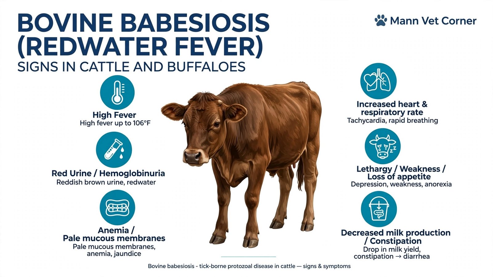
CBC results and interpretation
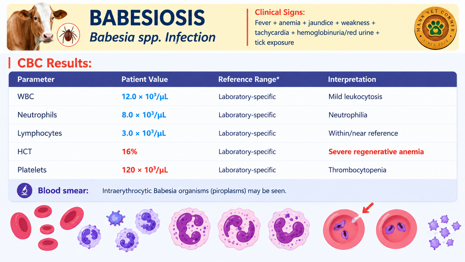
Table 10: Representative CBC in bovine babesiosis.
Blood smear: Intra-erythrocytic Babesia organisms (piroplasms) may be seen on a Giemsa-stained thin smear. Capillary blood from the ear or tail tip often shows more B. bovis parasites than jugular blood.
Why the CBC looks this way
Babesia parasites invade and destroy red blood cells. The result is intravascular hemolysis, which produces the hallmark combination of falling hematocrit, hemoglobinuria (red urine) and jaundice. The hematocrit of 16% shows how quickly oxygen-carrying capacity can collapse. The mild neutrophilia reflects systemic inflammation, and platelets fall because of consumption and inflammatory activation.
Regeneration deserves a note in cattle. Unlike dogs, cattle rarely release reticulocytes into the bloodstream even when the marrow is responding, so regeneration is judged by anisocytosis, polychromasia and basophilic stippling on the smear. It also takes several days to appear, so an acutely severe anemia may look poorly regenerative at first.
Quick decision guide
- Low WBC + low neutrophils + low lymphocytes, with vomiting and bloody diarrhea in a young cat or dog: think parvoviral disease (panleukopenia or CPV).
- Low WBC with lymphopenia, respiratory and neurological signs in an unvaccinated dog: think canine distemper.
- Low WBC with mouth lesions, eye and nose discharge and diarrhea in a goat or sheep: think PPR and notify authorities.
- High WBC with neutrophilia, persistent fever and effusion in a young cat: consider FIP.
- High WBC with neutrophilia, fever and abscessing jaw lymph nodes in a horse: think strangles.
- Severe anemia, low platelets and red urine in tick-exposed cattle: think babesiosis and look for parasites on the smear.
Frequently Asked Questions About CBC in Animal Disease Diagnosis
What is a CBC in animals?
A complete blood count (CBC) is a laboratory test that measures the cells in a blood sample: red blood cells (and hematocrit or PCV), white blood cells with a differential count (neutrophils, lymphocytes, monocytes, eosinophils, basophils), and platelets. Veterinarians use it to detect anemia, infection, inflammation, immune suppression and clotting-cell problems in dogs, cats, horses, cattle, sheep and goats.
Can a CBC diagnose parvovirus in dogs?
A CBC cannot confirm canine parvovirus on its own, but severe leukopenia with neutropenia and lymphopenia in a young puppy with vomiting and bloody diarrhea strongly supports it. Confirmation requires a fecal antigen test or PCR.
Why is the white blood cell count low in parvo and panleukopenia?
Parvoviruses destroy rapidly dividing cells, including bone marrow precursors of white blood cells and lymphoid tissue. At the same time, damaged intestines allow bacteria and inflammatory cells to be lost or consumed, which pushes neutrophil and lymphocyte counts down.
What does a high white blood cell count with neutrophilia mean in animals?
Neutrophilia usually signals inflammation or bacterial infection, such as strangles in horses or feline infectious peritonitis in cats. It can also be caused by stress, corticosteroids or tissue damage, so it must be interpreted with clinical signs and the blood smear.
Why is lymphopenia common in viral diseases?
Many viruses, including canine distemper virus, parvoviruses and PPR virus, infect or deplete lymphoid tissue, and stress hormones also redistribute lymphocytes. The result is a low lymphocyte count and a weakened immune response.
Which CBC findings suggest babesiosis in cattle?
Severe anemia with a very low hematocrit, thrombocytopenia and a mild neutrophilia in a febrile animal with red urine and tick exposure point toward bovine babesiosis. A Giemsa-stained blood smear showing intra-erythrocytic parasites, or PCR, confirms the diagnosis.
Can a normal CBC rule out disease?
No. Many diseases, including early infections, neurological disease and some parasitic infections, can leave the CBC within reference intervals. A normal result lowers suspicion for some conditions but never replaces clinical examination and specific tests.
How often should a CBC be repeated in a sick animal?
In hospitalized patients with rapidly changing diseases such as parvovirus enteritis, clinicians commonly repeat the CBC or PCV and total protein every day or as clinical status demands. The interval is a veterinary decision based on severity and response to treatment.
Do reference ranges differ between species?
Yes. Dogs, cats, horses, cattle, sheep and goats have different normal ranges for white cells, hematocrit and platelets, and values also vary with age, breed, pregnancy and the analyzer used. Always compare results with the reference intervals of the laboratory that ran the sample.
Is CBC testing useful for livestock health?
Yes. In cattle, buffalo, sheep and goats, a CBC helps to separate tick-borne hemolytic disease from viral or bacterial illness, to grade anemia and to monitor response to treatment. In large herds it is best combined with a blood smear and targeted PCR or serology.
Conclusion: Let the Pattern Guide the Diagnosis
The complete blood count is one of the most powerful first-line tests in veterinary medicine because it reveals how the whole immune and hematopoietic system is responding to disease. A low white cell count with neutropenia and lymphopenia shouts viral disease. A high white count with neutrophilia points toward bacterial infection or inflammation. A collapsing hematocrit with low platelets in a tick-exposed bovine demands a blood smear for Babesia.
Yet the CBC is a compass, not a map. It tells the clinician which direction to go, and PCR, antigen tests, culture, serology and careful microscopy show exactly where they have arrived. Read patterns rather than isolated numbers, respect species differences, account for dehydration and stress, and always look at the smear.
If your animal has a CBC report that worries you, or if you manage a herd with sudden fever, anemia or diarrhea, contact a licensed veterinarian immediately. Early diagnosis and supportive care often make the difference between recovery and loss, especially in young animals and in transboundary diseases such as PPR.
For more veterinary diagnostics guides, explore Cattle Daily’s library of articles on cattle, small ruminant, equine and companion animal health.
