Dive into Placental Diversity in Mammals: A Comprehensive Review and uncover the awe-inspiring world of the placenta, nature’s ultimate life-support system! This remarkable organ, a cornerstone of mammalian reproduction, acts as a dynamic bridge between mother and baby, orchestrating nutrient exchange, gas transfer, waste removal, and hormone production with breathtaking precision. Why should you read Placental Diversity in Mammals: A Comprehensive Review? To unveils the stunning variety of placental structures across species, from the sprawling diffuse placenta of horses to the compact discoid placenta of humans, each placenta shaped by millions of years of evolutionary genius. This article offers veterinary professionals, students, and curious minds a captivating journey through Placental Diversity in Mammals: A Comprehensive Review, blending detailed science with the thrill of discovery. You’ll explore how these adaptations reflect unique reproductive strategies, gain insights into their roles in domestic and wild mammals, and marvel at the placenta’s evolutionary significance—making this a must-read for anyone passionate about life’s beginnings!
Functional Roles of the Placenta
The placenta is a multifunctional organ performing several physiological roles essential for gestational success:
Nutrient and Gas Exchange
Facilitates bidirectional transfer of oxygen, carbon dioxide, glucose, amino acids, and lipids via specialized transport mechanisms.
Implements countercurrent exchange systems in some species (e.g., ruminants) to maximize efficiency.
Endocrine Regulation
Secretes progesterone, estrogen, placental lactogen, and relaxin to maintain pregnancy, modulate maternal metabolism, and prepare for parturition.
In equids and primates, the placenta assumes full progesterone production after corpus luteum regression.
Immunological Modulation
Establishes immune tolerance to prevent maternal rejection of the fetal allograft.
Expresses FAS ligand and indoleamine 2,3-dioxygenase (IDO) to suppress cytotoxic T-cell activity.
Barrier Function
Limits transfer of pathogens and xenobiotics via selective membrane transporters (e.g., P-glycoprotein).
Note: Lipid-soluble compounds (e.g., some antibiotics, steroids) readily cross, necessitating cautious drug use in pregnancy.
Structural Organization of the Placenta
The placenta comprises fetal and maternal components arranged in species-specific configurations:
1. Fetal Components
Chorion: Outer membrane giving rise to chorionic villi, the primary exchange units. Villous structure (e.g., branched vs. unbranched) dictates functional efficiency.
Amnion: Avascular inner membrane secreting amniotic fluid, which cushions the fetus and prevents adhesions.
2. Maternal Components
Decidua: Modified endometrium that interacts with chorionic villi. Decidual cells secrete:
Prostaglandins (initiate parturition)
Cytokines (regulate trophoblast invasion)
Extracellular matrix proteins (anchor placenta)
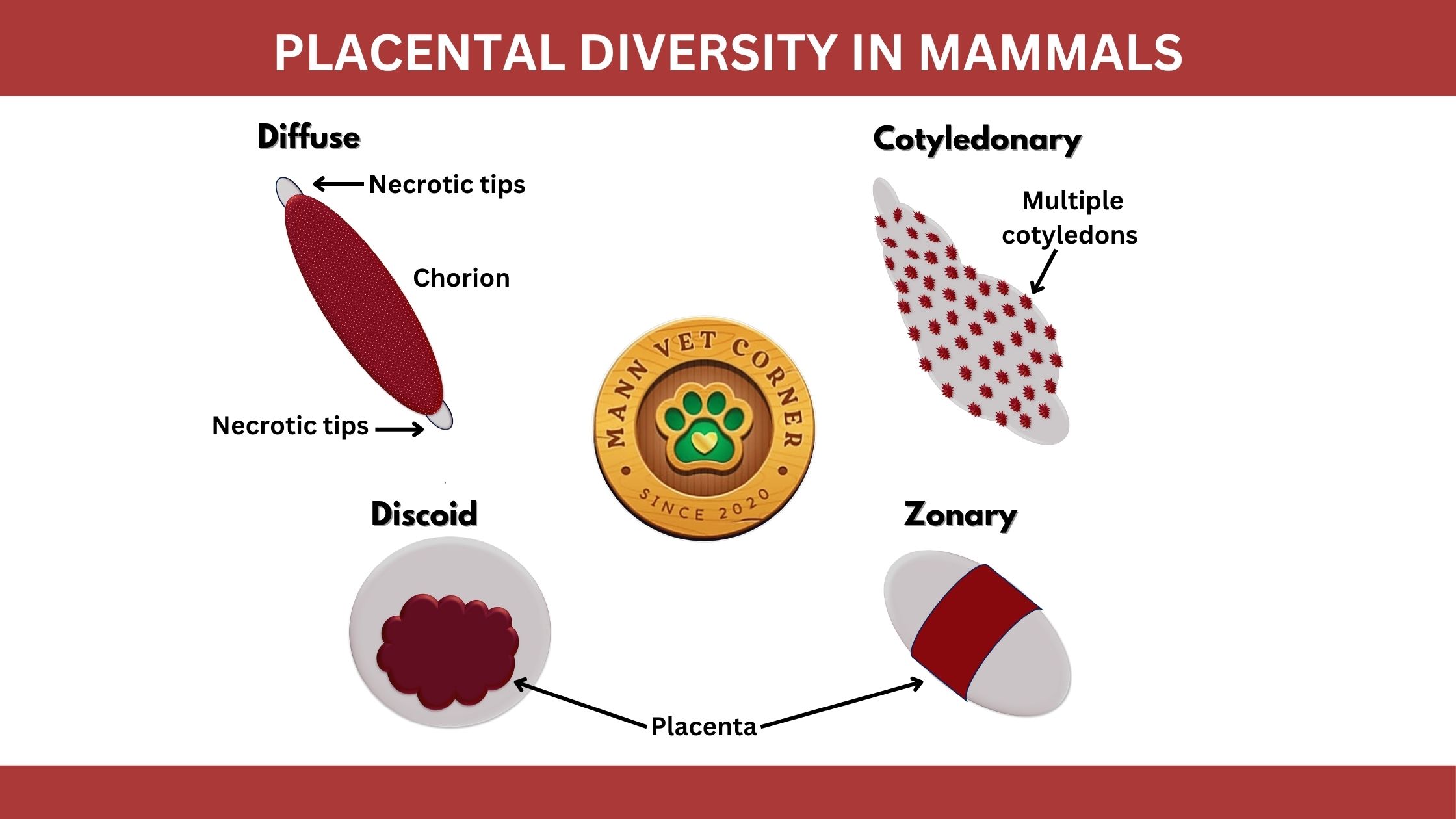
Placental Classification Criteria
Placentas are categorized by:
Shape and Contact Area (Gross morphology)
Histological Layers (Degree of invasiveness)
Duration of Function (Short vs. long gestation species)

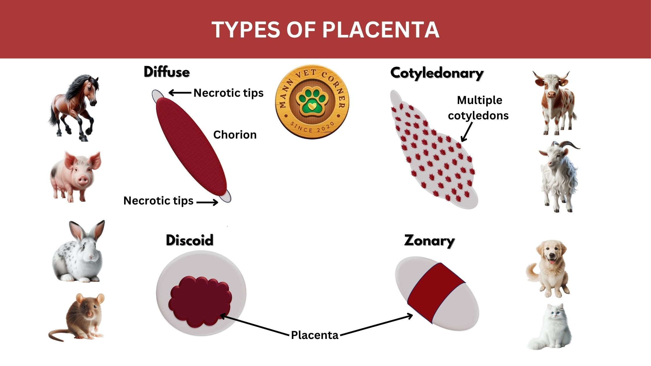
Comparative Analysis of Placental Types
1. Diffuse Placenta
The diffuse placenta is like a sprawling network. Its chorionic villi spread evenly across the uterine wall, creating a vast contact zone for nutrient and gas exchange. This setup maximizes surface area, ensuring steady delivery of oxygen and nutrients. Imagine a lush carpet of connections! Found in animals like pigs, horses, and whales, the diffuse placenta suits species with large, spread-out uterine surfaces. Interestingly, this type allows for prolonged pregnancies, as seen in horses, which carry their foals for nearly a year. The diffuse placenta’s design is less invasive, maintaining a looser connection with the mother’s tissues, which minimizes uterine damage during birth.
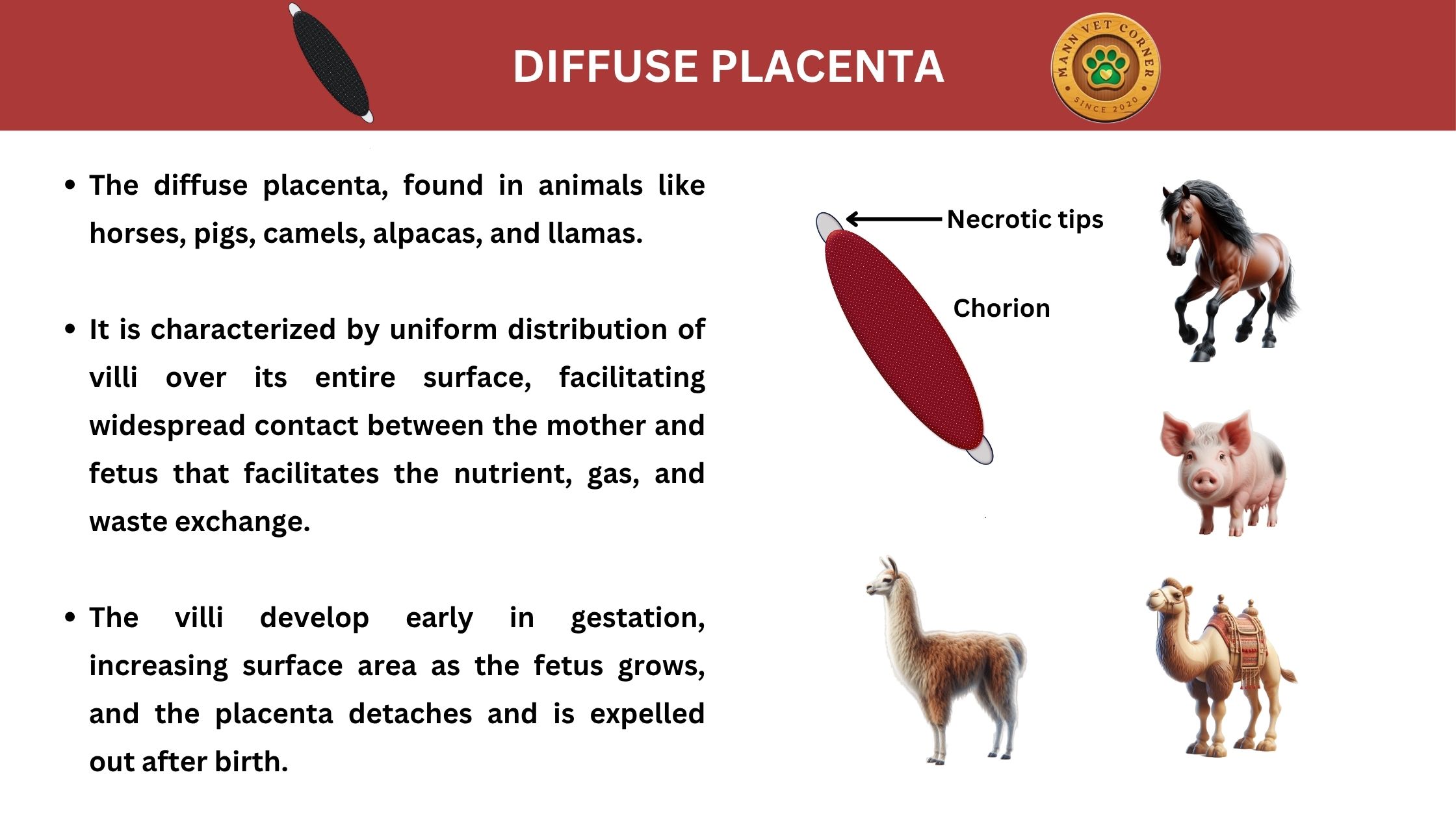
Morphology: Chorionic villi uniformly distributed across the chorionic sac (e.g., pigs, horses).
Histology: Epitheliochorial (least invasive; maternal epithelium remains intact).
Functional Adaptations:
Ideal for species with long gestations (e.g., equids: 340 days).
Allows fetal mobility, reducing umbilical cord torsion risk.
Clinical Relevance:
Retained placenta is rare due to weak adhesion.
In mares, endometrial cups (specialized trophoblast cells) secrete eCG to maintain pregnancy.
2. Cotyledonary Placenta
The cotyledonary placenta is a marvel of localized efficiency. It features multiple attachment points called cotyledons, which interlock with uterine caruncles to form structures known as placentomes. These placentomes act like mini-factories for nutrient and gas exchange, offering precision and high efficiency. Ruminants like cattle, sheep, and goats rely on this type, which supports their relatively large offspring. Did you know a cow’s placenta can have up to 100 cotyledons? This intricate system allows ruminants to nourish multiple fetuses, as seen in sheep with twin or triplet lambs. The cotyledonary placenta’s design also minimizes maternal blood loss during delivery, a key adaptation for grazing animals that need to recover quickly.
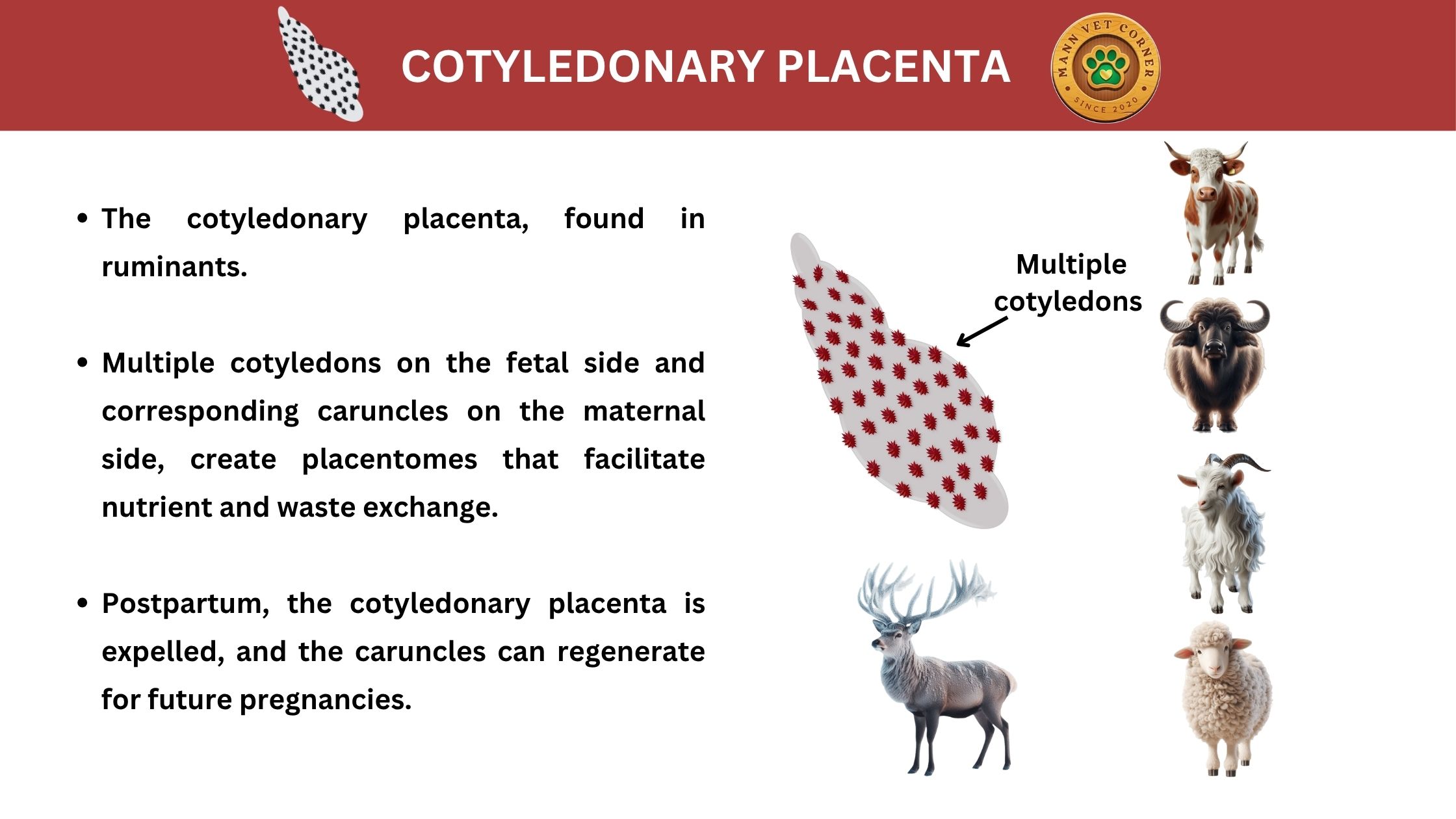
Morphology: Discrete cotyledons (fetal villi clusters) interlock with uterine caruncles to form placentomes (ruminants).
Histology: Synepitheliochorial (binucleate trophoblast cells fuse with uterine epithelium).
Functional Adaptations:
High-efficiency nutrient transfer supports rapid fetal growth (e.g., bovine fetuses gain ~1 kg/day late gestation).
Progesterone synthesis shifts to placentomes after day 150 in cattle.
Clinical Relevance:
Retained fetal membranes occur if placentomes fail to detach (risk factors: selenium deficiency, dystocia).
Neospora caninum exploits binucleate cells for vertical transmission.
3. Zonary Placenta
The zonary placenta is a striking band of connection. It forms a belt-like zone around the placenta’s midsection, creating a specialized area for blood exchange between mother and fetus. This type is common in carnivores like dogs, cats, and bears, whose active lifestyles demand efficient fetal nourishment. The zonary structure is highly vascular, ensuring rapid transfer of oxygen and nutrients to support fast-growing embryos. Fun fact: in dogs, the zonary placenta’s greenish hue comes from a pigment called uteroferrin, which aids iron transport to the fetus! This placenta type balances efficiency with flexibility, allowing carnivores to adapt to varied litter sizes and gestation periods.
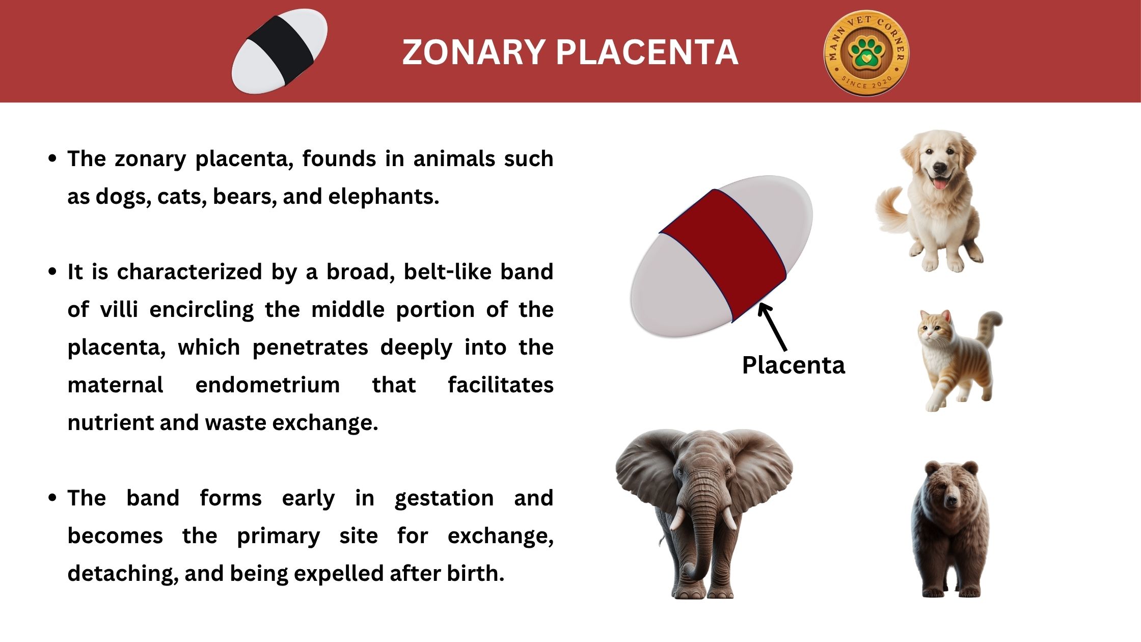
Morphology: Villi form a circumferential band (e.g., carnivores—dogs, cats).
Histology: Endotheliochorial (trophoblasts contact maternal endothelium).
Functional Adaptations:
Hemophagous zones at band edges absorb iron from lysed maternal RBCs.
Efficient for short gestations (canine: 63 days; feline: 65 days).
Clinical Relevance:
Green discoloration from uteroferrin is normal at parturition.
Placental infarcts may indicate systemic hypertension.
4. Discoid Placenta
The discoid placenta is a compact powerhouse. Shaped like a disk or circle, it concentrates nutrient and gas exchange in a single, highly efficient area. Humans, rodents, and some primates rely on this type, which supports rapid fetal development. Its small size belies its incredible capacity—tiny villi in the discoid placenta create a massive surface area for exchange, rivaling a football field in some species! This type is deeply invasive, embedding firmly into the uterine wall, which ensures robust nutrient delivery but can complicate delivery. In humans, the discoid placenta’s efficiency supports the long gestation and complex brain development of newborns.
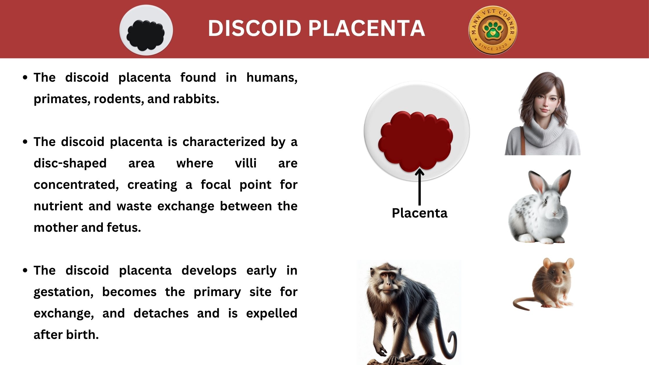
Morphology: Single disc-shaped exchange region (e.g., primates, rodents).
Histology: Hemochorial (most invasive; chorionic villi bathe in maternal blood).
Functional Adaptations:
Maximizes nutrient/waste exchange for encephalized fetuses.
Extravillous trophoblasts remodel spiral arteries to ensure high blood flow.
Clinical Relevance:
Preeclampsia stems from failed spiral artery remodeling.
Placenta accreta results from excessive trophoblast invasion.
Placental Expulsion: Species-Specific Variations
The third stage of labor involves placental detachment via:
Uterine contractions reducing placental site area.
Vasoconstriction preventing hemorrhage.
| Species | Expulsion Time | Key Notes |
|---|---|---|
| Bovine | 6–12 hours | Retention >12 hr requires treatment |
| Equine | 30–180 minutes | Rapid expulsion reduces infection risk |
| Canine | Immediate | Dams typically ingest placenta |
| Human | 30 minutes | Manual removal if >60 minutes |
Why Placental Research Matters in Veterinary Science
Improving Neonatal Survival
Understanding placental efficiency aids in managing intrauterine growth restriction (IUGR) in livestock.
Disease Transmission
Pathogens like Brucella and Listeria exploit placental tropism.
Toxicology
Placentas bioaccumulate environmental toxins (e.g., heavy metals in marine mammals).
Biotechnology
Placental stem cells show promise in regenerative medicine.
Conclusion: The Placenta as an Evolutionary Masterpiece
From the diffuse placenta of the mare to the hemochorial disc of the human, placental diversity reflects millions of years of adaptive fine-tuning. For veterinarians, recognizing these variations is crucial for diagnosing gestational disorders, optimizing neonatal care, and advancing reproductive therapeutics. Future research into placental genomics and immunology promises breakthroughs in both veterinary and human medicine.