
White blood cells (WBCs), or leukocytes, are critical components of the immune system in animals, playing essential roles in combating infections, mediating inflammation, and orchestrating adaptive immune responses. In veterinary medicine, the evaluation of WBCs through clinical pathology provides invaluable insights into an animal’s health status, aiding in the diagnosis and management of diseases. This article explores the characteristics, functions, and clinical significance of WBCs—specifically neutrophils (or heterophils in birds), eosinophils, basophils, monocytes, and lymphocytes (T and B cells)—across a range of animals, including domestic pets (dogs, cats), avian species (chickens, parrots), ruminants (cattle, sheep, goats), horses, and camels. By integrating hematological data and clinical pathology perspectives, we aim to provide a comprehensive understanding of WBCs in veterinary practice.
-
How WBCs vary across species (mammals, birds, and ruminants).
-
Key clinical differences in WBC responses to infections.
The Role of White Blood Cells in Animal Immunity
Leukocytes are a diverse group of cells that protect animals from pathogens, foreign substances, and abnormal cells. They are produced in the bone marrow (or equivalent hematopoietic tissues, such as the bursa of Fabricius in birds) and circulate in the blood and tissues. Each type of WBC has distinct morphological and functional characteristics, which vary across species due to evolutionary adaptations. In clinical pathology, the complete blood count (CBC) is the cornerstone for evaluating WBCs, providing quantitative data on total and differential leukocyte counts. These measurements, combined with morphological assessments, help veterinarians identify conditions such as infections, allergic reactions, stress, or neoplasia.
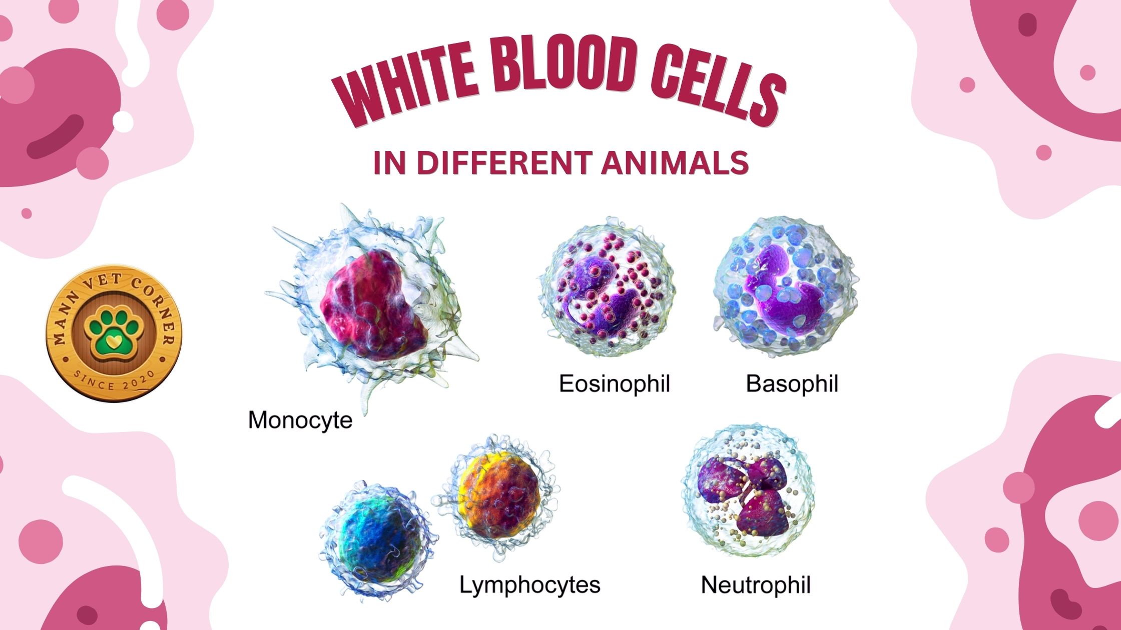
The differential WBC count categorizes leukocytes into five main types: neutrophils (heterophils in birds), eosinophils, basophils, monocytes, and lymphocytes. Reference ranges for these cells differ significantly among species, reflecting physiological adaptations to their environments and lifestyles. Below, we discuss each cell type, their reference ranges, and their clinical significance in dogs, cats, chickens, parrots, cattle, sheep, goats, horses, and camels, supported by a comprehensive table summarizing key data.
Neutrophils (Mammals) vs. Heterophils (Birds): The First Line of Defense
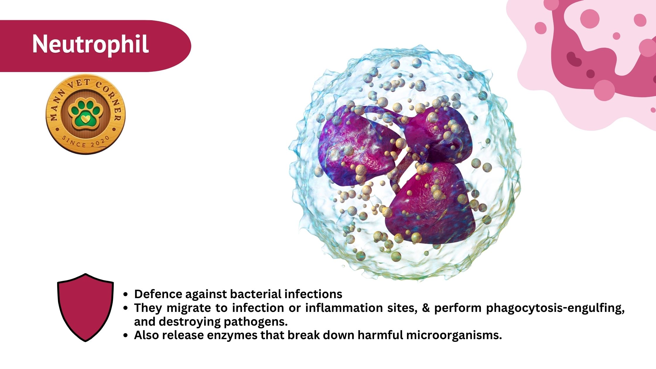
Neutrophils in mammals (and their avian equivalent, heterophils) are the primary responders to acute bacterial infections. These granulocytes are characterized by a multilobed nucleus and granular cytoplasm, which contains antimicrobial enzymes and peptides. In mammals, neutrophils excel at phagocytosis and intracellular killing, releasing reactive oxygen species and myeloperoxidase to neutralize pathogens. In contrast, avian heterophils rely more on extracellular killing mechanisms, such as heterophil extracellular traps (HETs), and lack myeloperoxidase, reflecting a distinct evolutionary strategy.
-
Neutrophils in Mammals (Dogs, Cats, Cows, Horses, Camels)
-
Primary Role: Rapid responders to bacterial infections.
-
-
Key Features:
-
Phagocytic (engulf and destroy bacteria).
-
Release reactive oxygen species (ROS) to kill pathogens.
-
Show toxic changes (vacuolation, Döhle bodies) in severe infections.
-
Species-Specific Differences:
-
Dogs & Cats: High neutrophil counts indicate bacterial infections (pyometra, abscesses).
-
Ruminants (Cows, Sheep, Goats): Normally have lower neutrophil percentages (lymphocytes dominate).
-
Camels: Unusually high baseline neutrophils (possibly due to harsh desert environments).
Heterophils in Birds (Chickens, Parrots, Eagles)
-
Primary Role: Defend against bacterial and fungal infections.
-
Key Differences from Neutrophils:
-
Do not contain myeloperoxidase (unlike mammalian neutrophils).
-
Use heterophil extracellular traps (HETs) to trap and kill pathogens.
-
Toxic heterophils (degranulation, abnormal shapes) indicate severe disease.
-
Clinical Significance in Birds:
-
Chickens: High heterophils → E. coli, Salmonella infections.
-
Parrots: Toxic heterophils → Chlamydia (psittacosis), aspergillosis.
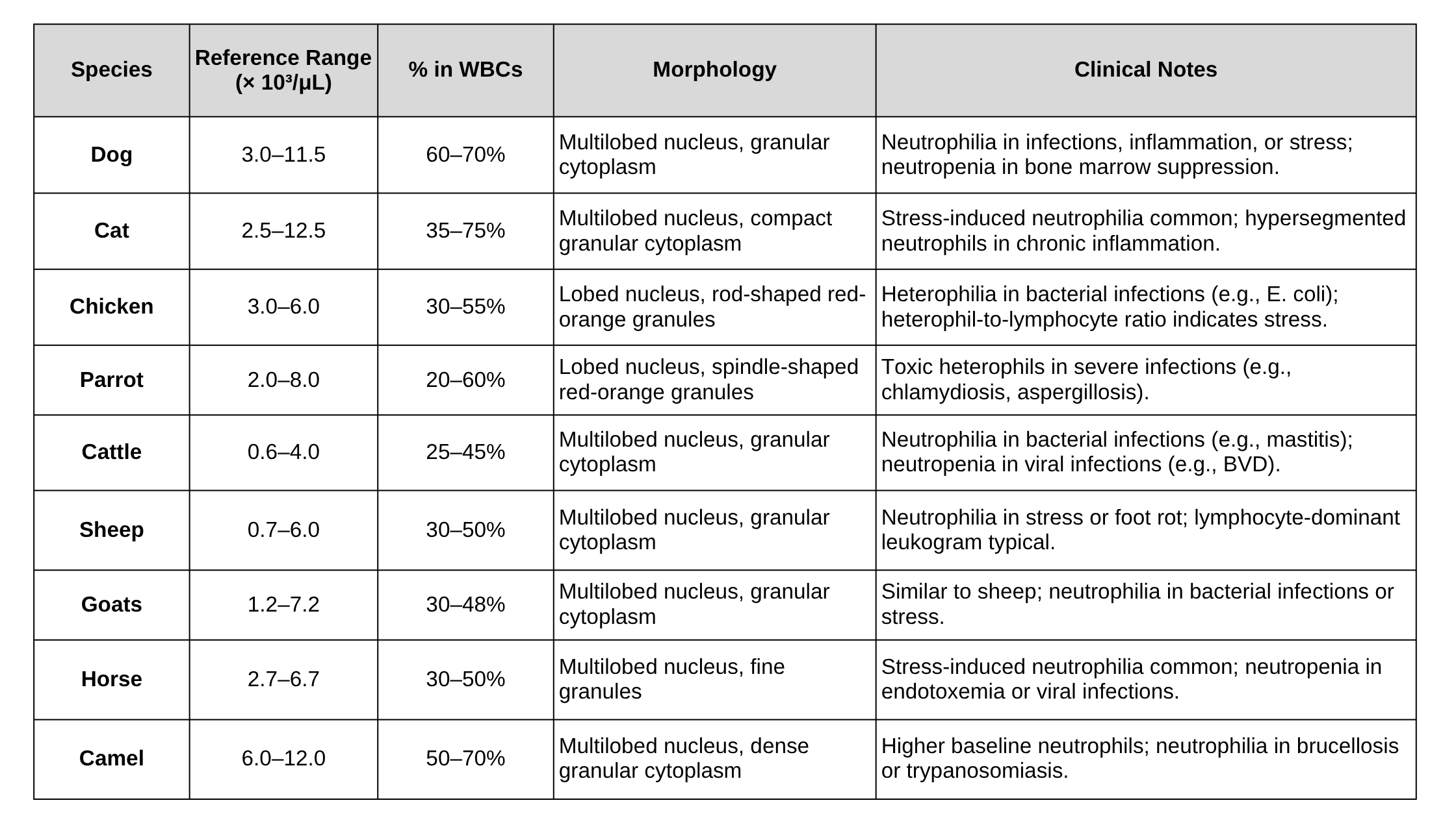
SUMMARIZE TABLE OF NEUTROPHIL/HETEROPHIL IN DIFFERENT ANIMALS
Eosinophils: Guardians Against Parasites and Allergies
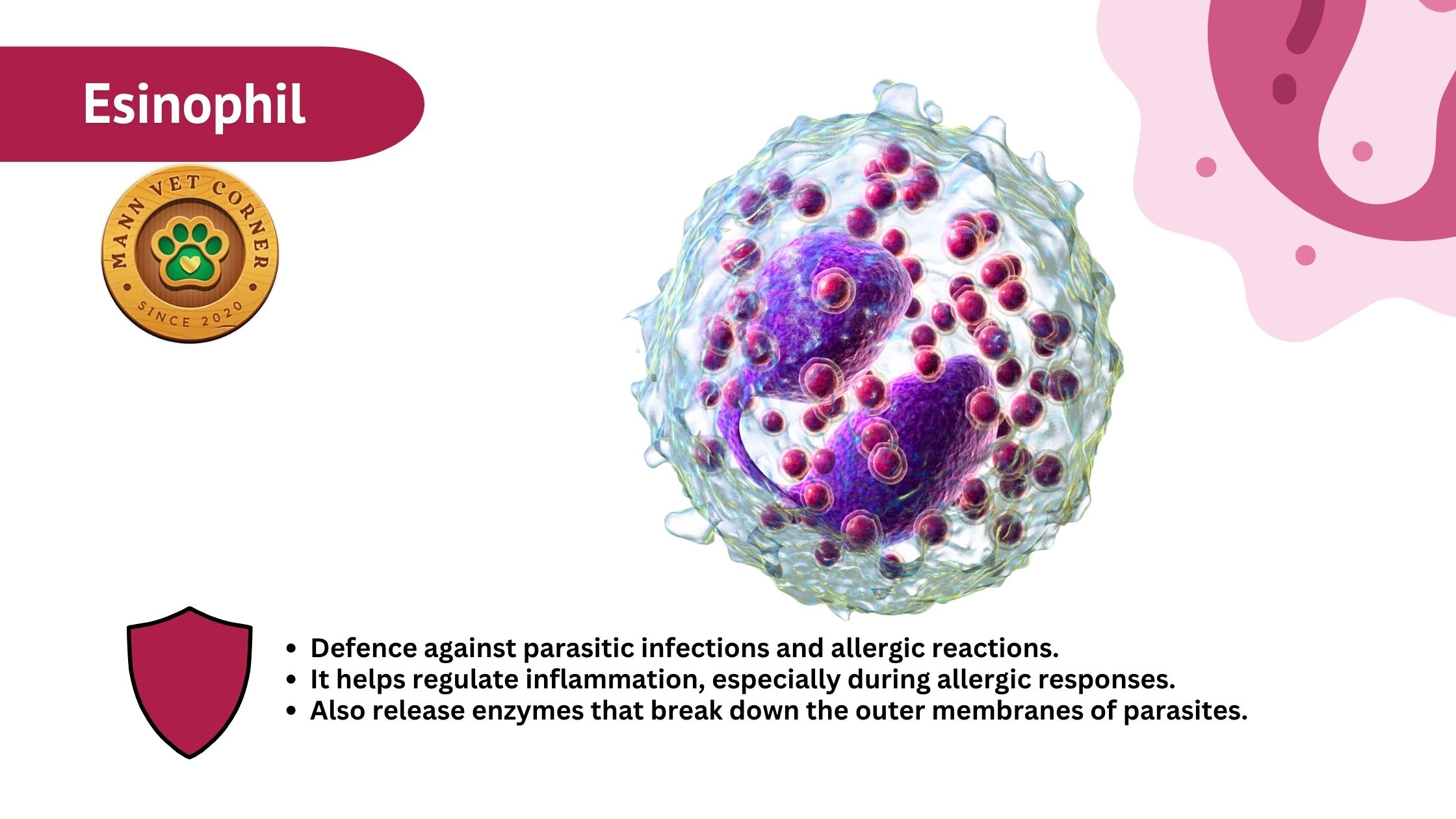
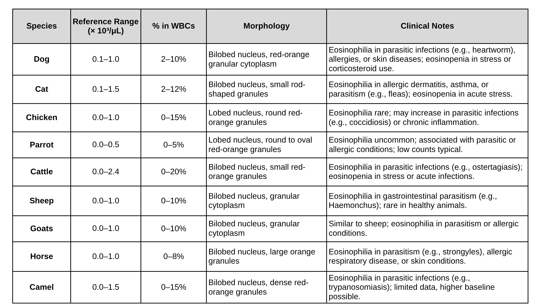
Eosinophils are granulocytes specialized in combating parasitic infections and mediating allergic responses. They are characterized by a bilobed nucleus and red-orange granules containing major basic protein and other antiparasitic molecules. Eosinophils are typically present in low numbers but increase significantly in specific conditions.
 Function:
-
Combat parasitic infections (worms, protozoa).
-
Involved in allergic reactions (asthma, skin allergies).
Species Variations:
| Animal | Eosinophil Response | Common Conditions |
|---|---|---|
| Dogs | High in heartworm, flea allergies | Eosinophilic dermatitis |
| Cats | Elevated in asthma, flea infestations | Feline eosinophilic granuloma |
| Sheep & Goats | Spike with Haemonchus (barber pole worm) | Parasitic gastroenteritis |
| Horses | Increase in strongyle worm infections | Larval cyathostominosis |
| Birds | Rare, but seen in coccidiosis | Intestinal parasite infections |
Note: Eosinopenia (low eosinophils) often occurs during stress or steroid therapy.
SUMMARIZE TABLE OF EOSINOPHILS IN DIFFERENT ANIMALS
Basophils: The Elusive Mediators
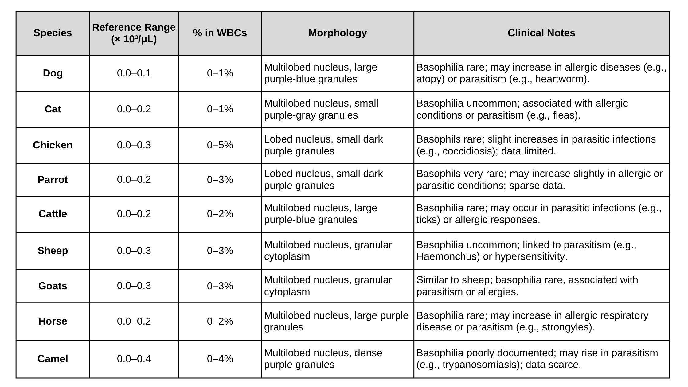
Basophils are the least abundant leukocytes, characterized by a multilobed nucleus and large purple-blue granules containing histamine and heparin. They play roles in allergic and parasitic responses but are rarely elevated, making their clinical significance challenging to assess.
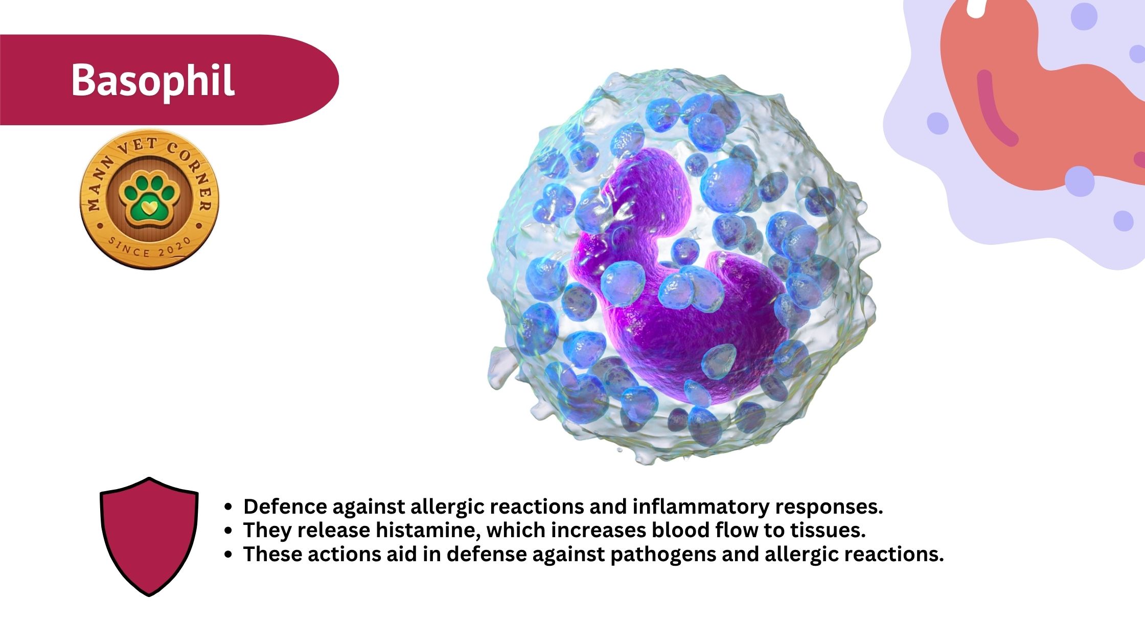 Function:
-
Release histamine (allergic responses).
-
Assist in parasite defense (especially in ruminants).
Key Points:
-
Low numbers in most animals (<1% of WBCs).
-
Basophilia (high basophils) is uncommon but seen in:
-
Dogs & Cats: Allergies, heartworm disease.
-
Ruminants (Cows, Sheep): Tick-borne diseases.
-
Birds: Occasionally in severe parasitism.
-
SUMMARIZE TABLE OF BASOPHILS IN DIFFERENT ANIMALS
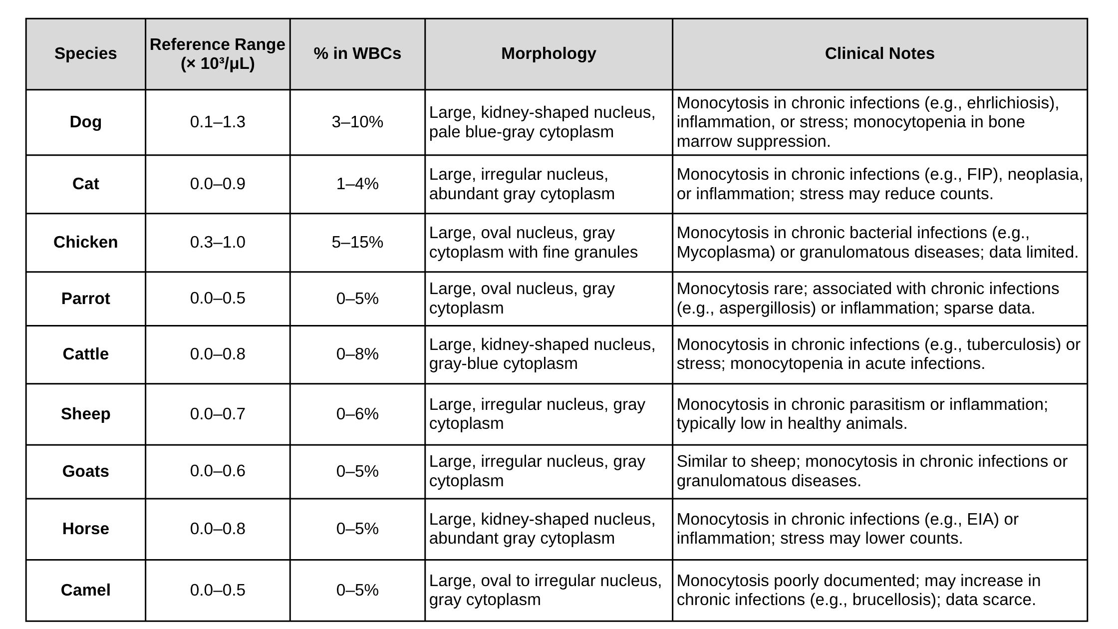
Monocytes: The Sentinels of Chronic Inflammation
Monocytes are large leukocytes with kidney-shaped or irregular nuclei and abundant gray cytoplasm. They differentiate into macrophages or dendritic cells in tissues, playing roles in phagocytosis, antigen presentation, and chronic inflammation.
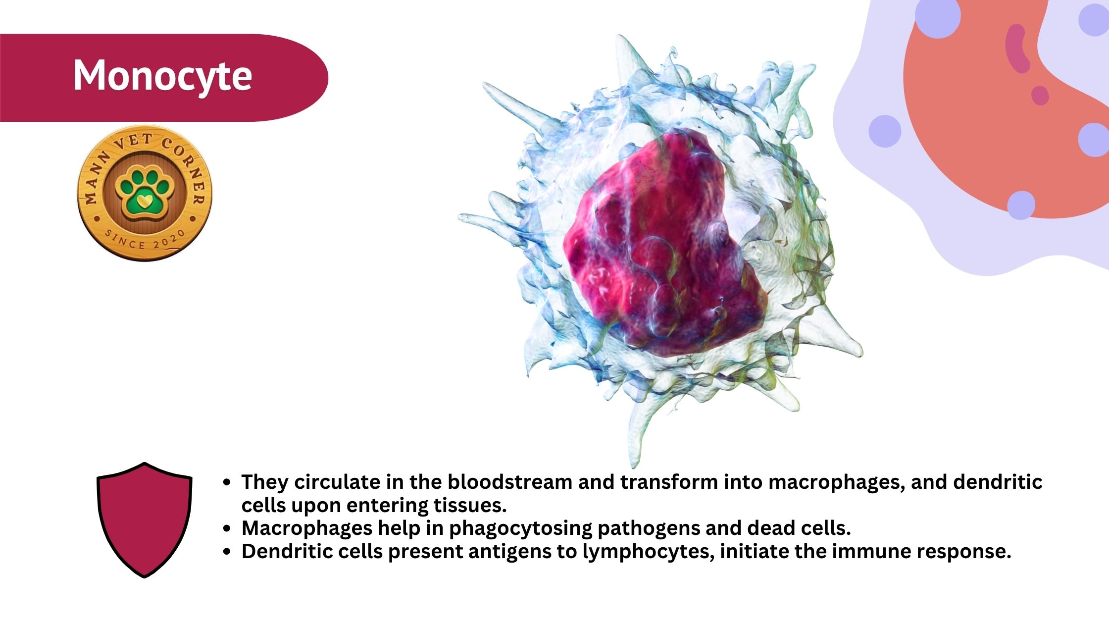 Function:
-
Differentiate into macrophages (clean up dead cells, chronic infections).
-
Present antigens to lymphocytes (adaptive immunity).
Clinical Significance:
| Animal | Monocytosis (High Monocytes) Indicates |
|---|---|
| Dogs | Ehrlichiosis, chronic inflammation |
| Cats | Feline Infectious Peritonitis (FIP) |
| Cows | Tuberculosis, brucellosis |
| Horses | Equine Infectious Anemia (EIA) |
| Birds | Aspergillosis, mycobacteriosis |
Note: Immature monocytes (left shift) suggest severe infections.
SUMMARIZE TABLE OF MONOCYTES IN DIFFERENT ANIMALS
Lymphocytes: Adaptive Immunity Specialists
Lymphocytes, comprising T cells (cell-mediated immunity) and B cells (humoral immunity), are critical for adaptive immune responses. They have a small, round nucleus with a thin rim of cytoplasm. Ruminants and avian species often have lymphocyte-dominant leukograms, reflecting their immune system priorities.
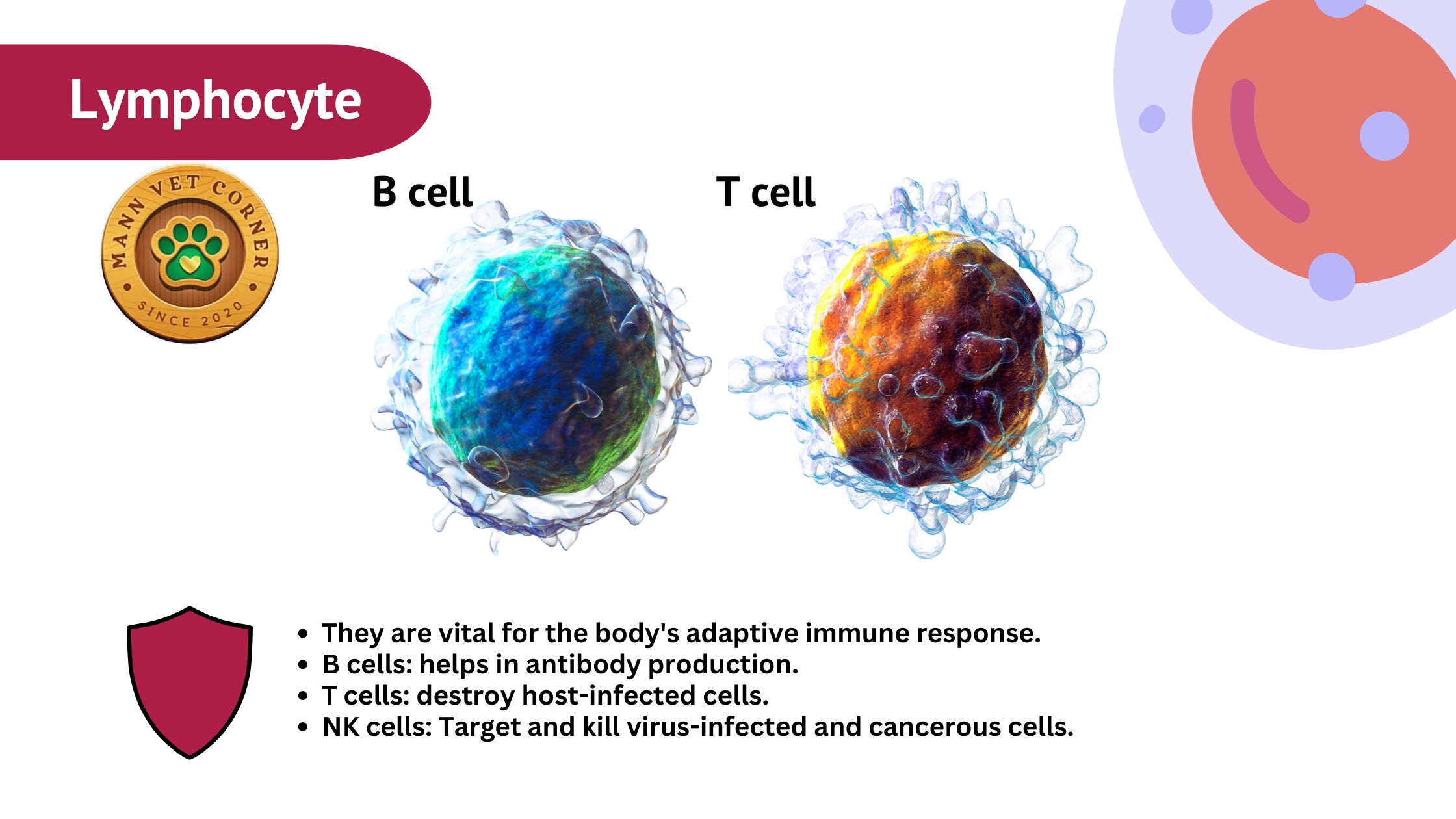 Function:
-
B cells: Produce antibodies (humoral immunity).
-
T cells: Direct cell-mediated immunity (kill infected cells).
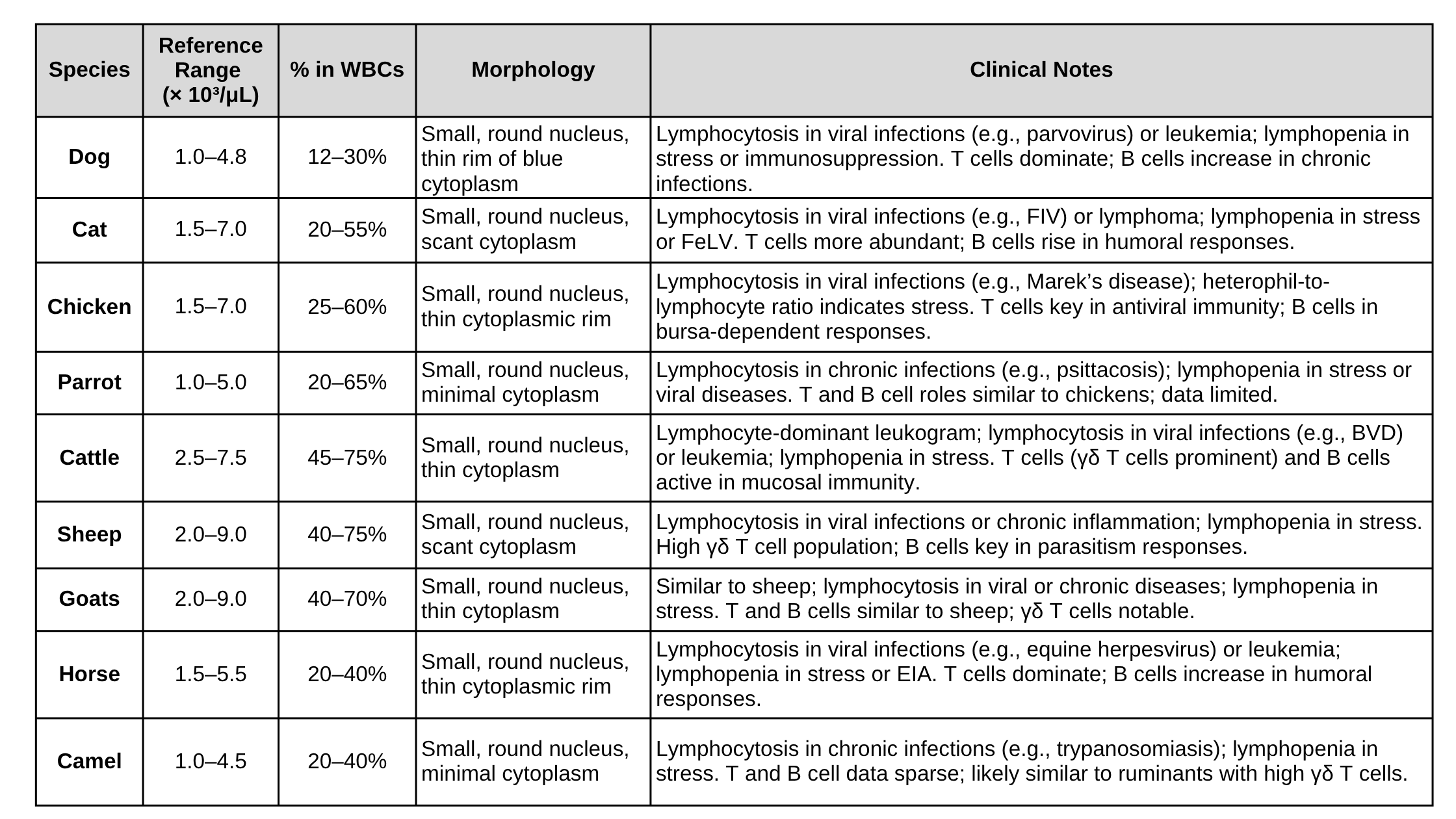
Species-Specific Patterns: Below table gives brief species- specific patterns of lymphocyte difference
| Animal | Lymphocyte Dominance? | Key Facts |
|---|---|---|
| Dogs & Cats | No (neutrophils dominate) | Lymphocytosis = viral infections (parvovirus, FeLV) |
| Ruminants (Cows, Sheep, Goats) | Yes! (45-75% of WBCs) | High γδ T cells (unique immune defense) |
| Birds (Chickens, Parrots) | Yes (25-60%) | Marek’s disease (viral lymphoma) destroys lymphocytes |
| Horses | Moderate (20-40%) | Lymphopenia in equine herpesvirus |
| Camels | Lower (20-40%) | Still under research |
SUMMARIZE TABLE OF LYMPHOCYTES IN DIFFERENT ANIMALS
Clinical Pathology Applications
The CBC is a powerful diagnostic tool in veterinary medicine, but interpreting WBC counts requires context. Stress leukograms (neutrophilia, lymphopenia, eosinopenia) are common in cats, horses, and camels, necessitating careful history-taking. Left shifts, toxic changes, or atypical lymphocytes on blood smears provide additional diagnostic clues. For example, toxic heterophils in parrots suggest severe infections, while monocytosis in cattle may point to tuberculosis. Species-specific reference ranges are crucial, as camels have higher baseline neutrophils, and ruminants have lymphocyte-dominant profiles.
Conclusion
White blood cells are indispensable for understanding animal health, and their evaluation through clinical pathology is a cornerstone of veterinary diagnostics. By analyzing neutrophil, eosinophil, basophil, monocyte, and lymphocyte counts, veterinarians can diagnose a wide range of conditions, from acute infections to chronic diseases and stress responses. Species-specific differences, such as the high neutrophil counts in camels or lymphocyte dominance in ruminants, highlight the need for tailored reference ranges and careful interpretation. As research advances, particularly for understudied species like camels, our ability to leverage WBC data in clinical practice will continue to improve, enhancing animal care across diverse veterinary contexts.
