Blood collection and preservation are critical processes in medical and veterinary diagnostics, research, and treatment. Whether for routine blood tests, emergency diagnostics, transfusions, or specialized laboratory analysis, preventing blood from clotting is essential to maintain sample integrity and usability. Anticoagulants, often referred to as ”blood thinners” in clinical settings, play a vital role in this process by inhibiting the blood’s natural clotting mechanisms.
These agents ensure that blood samples remain in a liquid state for accurate testing, safe storage, and reliable diagnostic results. Without proper anticoagulation, blood samples would quickly solidify, making laboratory analysis impossible and potentially compromising patient care. This comprehensive guide explores the intricate science of blood clotting, the critical role of anticoagulants in blood collection, and its mechanisms.
By understanding these fundamental concepts, healthcare professionals can better appreciate how anticoagulant systems support accurate medical procedures and improve patient outcomes. This knowledge is essential for laboratory technicians, vets, field veterinarians, phlebotomists, and other animal workers involved in blood collection and processing.
Coagulation Mechanism: How Blood Clots Form
Blood clotting, or coagulation, is a remarkably complex physiological process that prevents excessive bleeding when blood vessels are injured. This sophisticated biological mechanism involves a precisely orchestrated cascade of events that transforms liquid blood into a solid clot, effectively sealing wounds and maintaining vascular integrity. Understanding this coagulation process is fundamental to appreciating how anticoagulants function and why they are essential in laboratory medicine.
The Coagulation Cascade: A Three-Stage Process
The coagulation process involves three main stages that work in concert:
1. Vascular Response When a blood vessel is damaged, the vessel immediately constricts (vasoconstriction) to reduce blood flow and minimize blood loss. This initial response provides the body with crucial time to initiate more permanent hemostatic mechanisms.
2. Platelet Activation Platelets, small cell fragments circulating in the blood, rapidly adhere to the injury site through interaction with exposed collagen and von Willebrand factor. These activated platelets form a temporary plug and release chemical signals that attract additional platelets while simultaneously initiating the clotting cascade.
3. Clot Formation The clotting cascade activates a series of proteins called clotting factors in a precise sequence of enzymatic reactions. This cascade ultimately leads to the conversion of fibrinogen (a soluble plasma protein) into fibrin, creating an insoluble mesh that reinforces the platelet plug to form a stable, definitive clot.

Key Components of the Coagulation System
Clotting Factors The coagulation system involves numerous clotting factors, each designated by Roman numerals:
- Factor I (Fibrinogen): The soluble precursor to fibrin clot formation
- Factor II (Prothrombin): Converts to thrombin, the key enzyme in clot formation
- Factor III (Thromboplastin/Tissue Factor): Initiates the extrinsic coagulation pathway
- Factor IV (Calcium): Essential cofactor for multiple coagulation reactions
These proteins, primarily synthesized in the liver, require specific cofactors for activation and function.
Essential Cofactors
- Calcium Ions: Critical for stabilizing clotting factor complexes and enabling multiple steps in the coagulation cascade
- Vitamin K: Required for the post-translational modification of factors II, VII, IX, and X
- Phospholipids: Provide surfaces for coagulation factor complex assembly
Fibrinogen and Fibrin Conversion The final step involves thrombin converting soluble fibrinogen into insoluble fibrin strands. These strands create a three-dimensional mesh that traps blood cells, platelets, and plasma proteins, forming the definitive hemostatic plug.
When blood is collected for laboratory testing or storage, this natural clotting process must be completely inhibited to prevent sample solidification and ensure accurate analytical results.
Anticoagulants: Types, Mechanisms, and Applications
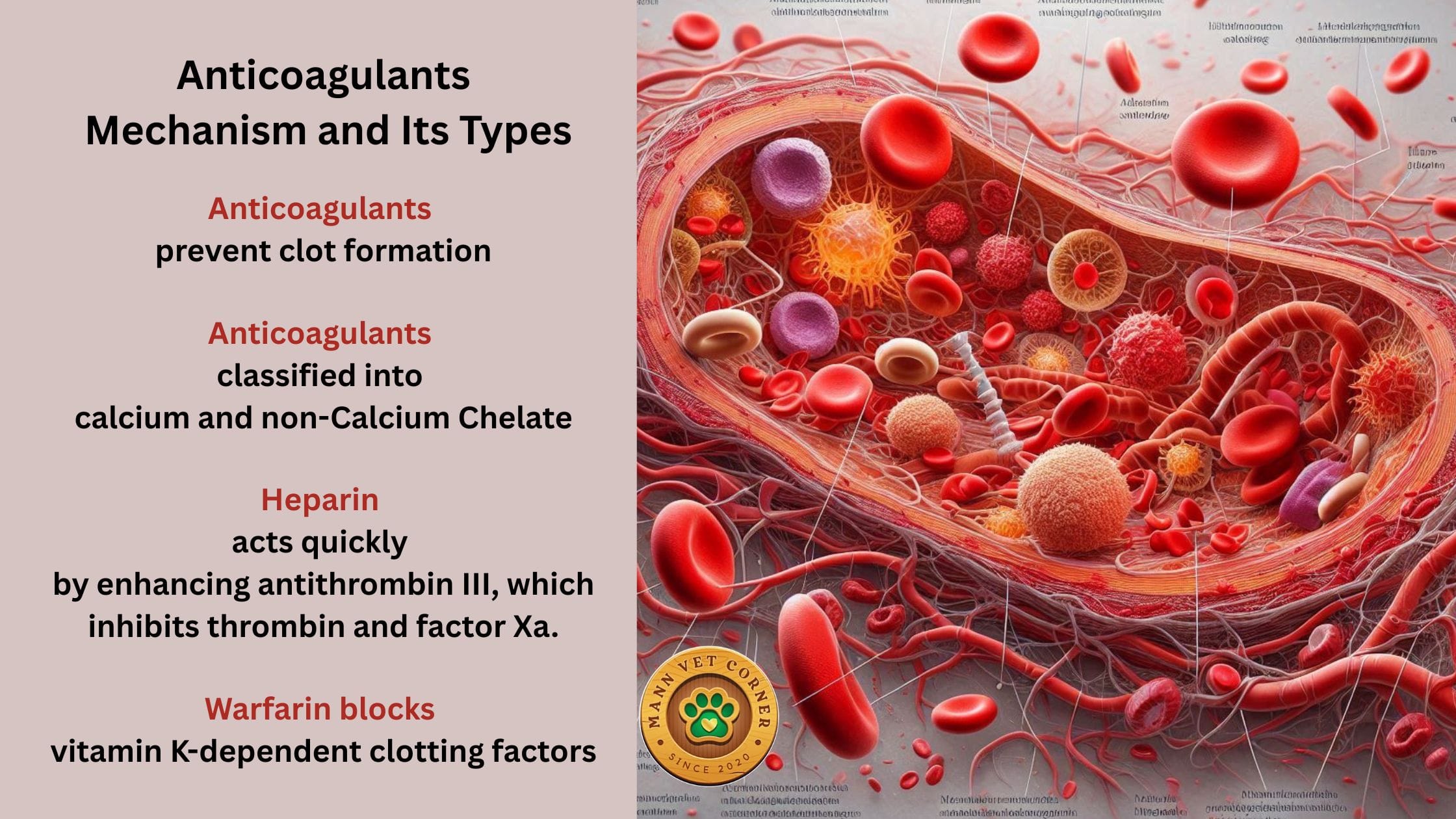
Anticoagulants are specialized substances designed to prevent blood coagulation through various mechanisms. In laboratory settings, they are incorporated into blood collection tubes to maintain samples in a liquid state for diagnostic testing. In clinical medicine, they are prescribed as therapeutic agents to prevent pathological clot formation in conditions such as atrial fibrillation, deep vein thrombosis, or pulmonary embolism.

How Anticoagulants Disrupt the Clotting Mechanism
Anticoagulants prevent blood clot formation by targeting key steps in the coagulation cascade:
- 🧪 Calcium Inhibition: Calcium ions act as essential cofactors in activating clotting enzymes. Agents like EDTA or citrate bind calcium, halting the cascade at its early stages.
- 🧬 Thrombin Blockade: Thrombin converts fibrinogen into fibrin, the structural mesh of clots. Drugs like heparin or direct thrombin inhibitors neutralize thrombin, preventing fibrin formation.
- 🧫 Platelet Aggregation & Fibrin Formation: Some anticoagulants also impair platelet activation and aggregation (e.g., aspirin, clopidogrel), reducing the cellular component of clots. Others inhibit fibrin polymerization, weakening clot stability.
Anticoagulants can be broadly classified into two major categories based on their mechanism of action: calcium chelators and non-calcium chelators.
1. Calcium Chelating Anticoagulants
Calcium chelators function by sequestering calcium ions in blood, which are absolutely essential for multiple steps in the coagulation cascade. By removing bioavailable calcium, these anticoagulants effectively prevent clotting factor activation and halt clot formation.
EDTA (Ethylenediaminetetraacetic Acid)
Mechanism: EDTA forms extremely stable chelate complexes with divalent cations, particularly calcium and magnesium. This strong binding affinity effectively removes these ions from the coagulation process.
Variants:
- K₂EDTA (Dipotassium EDTA): Most commonly used in blood collection
- K₃EDTA (Tripotassium EDTA): Alternative formulation with similar properties
- Na₂EDTA (Disodium EDTA): Less commonly used variant
Applications:
- Hematology testing (Complete Blood Count, blood films)
- Cell morphology preservation
- Molecular genetic studies (DNA/RNA extraction)
- Flow cytometry applications
Advantages: Excellent cell preservation, prevents platelet clumping, maintains cellular morphology
Limitations: Completely inhibits coagulation, making it unsuitable for coagulation studies
Sodium Citrate
Mechanism: Forms reversible chelate complexes with calcium ions, allowing controlled anticoagulation that can be overcome by adding excess calcium back to the sample.
Applications:
- Coagulation studies (PT, aPTT, fibrinogen assays)
- Blood banking and plasma preservation
- Platelet function testing
Advantages: Reversible anticoagulation, excellent for coagulation testing, good plasma preservation
Limitations: Less effective for long-term cell preservation compared to EDTA
Citrate-Based Preservative Solutions
- ACD (Acid Citrate Dextrose): Contains citric acid, sodium citrate, and dextrose for enhanced cell preservation
- CPD (Citrate Phosphate Dextrose): Includes phosphate buffer for improved pH stability
- CPDA (Citrate Phosphate Dextrose Adenine): Enhanced formulation with adenine for extended red blood cell storage
Oxalates
Mechanism: Form insoluble calcium oxalate precipitates, effectively removing calcium from the coagulation system.
Types:
- Ammonium Oxalate: Traditional anticoagulant with good preservation properties
- Potassium Oxalate: Commonly used for glucose testing
- Double Oxalate: Combination formulation for enhanced anticoagulation
Applications: Chemistry panels, glucose testing (often combined with fluoride)
Limitations: May interfere with cell morphology, less commonly used in modern practice

2. Non-Calcium Chelating Anticoagulants

Non-calcium chelating anticoagulants inhibit coagulation through mechanisms independent of calcium binding, typically by directly targeting specific clotting factors or enzymes in the coagulation cascade.
Heparin
Mechanism: Enhances the activity of antithrombin III, a natural anticoagulant protein that inhibits key clotting factors including thrombin (Factor IIa) and Factor Xa. Heparin acts as a cofactor, dramatically accelerating antithrombin’s inhibitory activity.
Types:
- Sodium Heparin: Most common formulation for laboratory use
- Lithium Heparin: Alternative salt form with similar properties
- Unfractionated Heparin (UFH): Full-length heparin molecules
- Low Molecular Weight Heparin (LMWH): Smaller fragments with more predictable activity
Applications:
- Plasma-based chemistry testing
- Electrolyte analysis
- Therapeutic anticoagulation in clinical settings
Advantages: Rapid onset, suitable for both diagnostic and therapeutic applications
Limitations: Variable activity, potential for heparin-induced thrombocytopenia in clinical use
Warfarin and Vitamin K Antagonists
Mechanism: Inhibits vitamin K-dependent clotting factors (II, VII, IX, X) by interfering with the vitamin K recycling pathway in hepatocytes, preventing proper factor synthesis and activation.
Applications: Primarily therapeutic anticoagulation for long-term clot prevention
Advantages: Effective oral anticoagulant, reversible with vitamin K administration
Limitations: Requires regular monitoring, multiple drug and dietary interactions
Direct Oral Anticoagulants (DOACs)
Mechanisms:
- Direct Thrombin Inhibitors (e.g., dabigatran): Directly bind and inhibit thrombin
- Factor Xa Inhibitors (e.g., apixaban, rivaroxaban): Directly inhibit Factor Xa
Applications: Modern therapeutic anticoagulation with improved safety profiles
Advantages: Predictable pharmacokinetics, minimal monitoring requirements
Limitations: Higher cost, limited reversal options in emergencies
Conclusion
The sophisticated understanding and proper application of anticoagulants and blood collection systems represent fundamental cornerstones of modern laboratory medicine and diagnostic accuracy. The coagulation mechanism, with its intricate cascade of factors and cofactors, provides the foundation for understanding why different anticoagulants are necessary for various diagnostic applications. Calcium chelators like EDTA and sodium citrate serve essential roles in preserving cellular elements and enabling coagulation studies, while non-calcium chelators such as heparin provide unique advantages for specific plasma-based analyses.
👍👍