Introduction
The Complete Blood Count (CBC) is one of the most fundamental diagnosis tools in animal disease, providing critical insights into an animal’s health status through analysis of blood cellular components. This comprehensive blood test evaluates red blood cells (RBCs), white blood cells (WBCs), and platelets, offering rapid, cost-effective screening for a wide range of clinical guide conditions from infections to systemic diseases.
CBC serves multiple functions in veterinary practice:
- Screening tool for apparently healthy animals during routine examinations
- Diagnostic aid for sick patients presenting with clinical signs
- Monitoring tool for tracking disease progression and treatment response
- Emergency diagnostic providing immediate information for critical decisions
The test provides essential parameters including:
- Red Blood Cell Analysis: RBC count, hemoglobin, hematocrit, and red cell indices (MCV, MCH, MCHC)
- White Blood Cell Analysis: Total WBC count and differential (neutrophils, lymphocytes, monocytes, eosinophils, basophils)
- Platelet Analysis: Platelet count and morphology assessment
- Additional Parameters: Reticulocyte count, blood cell morphology, and various cellular indices

Understanding CBC interpretation requires knowledge of species-specific reference ranges, recognition of diagnostic patterns, and integration with clinical findings for accurate diagnosis and appropriate treatment decisions.
Case Study 1: Canine Parvovirus Infection
Disease Overview
Canine parvovirus (CPV) is a highly contagious viral infection that primarily affects puppies and unvaccinated dogs. The virus targets rapidly dividing cells, particularly in the gastrointestinal tract and bone marrow, leading to severe enteritis and immunosuppression.
Case Presentation
Patient: 4-month-old German Shepherd puppy
Clinical Signs: Severe bloody diarrhea, vomiting, lethargy, dehydration
Interpretation
The severe leukopenia with neutropenia is pathognomonic for parvovirus infection. The virus specifically attacks bone marrow precursor cells, causing profound immunosuppression. The apparently normal hematocrit in a dehydrated patient suggests underlying anemia that will become evident once hydration is restored.
Clinical Significance
- Emergency status: Requires immediate aggressive treatment
- Treatment protocol: IV fluid therapy, antibiotics for secondary infections, anti-emetics, nutritional support
- Prognosis: Guarded to poor, especially with severe neutropenia in young animals
- Management: Strict isolation protocols essential to prevent spread
Case Study 2: Feline Iron-Deficiency Anemia
Disease Overview
Iron-deficiency anemia in cats typically results from chronic blood loss, most commonly from gastrointestinal or urinary tract bleeding. The condition develops gradually as iron stores become depleted, leading to decreased hemoglobin synthesis and characteristic microcytic anemia.
Case Presentation
Patient: 6-year-old domestic shorthair cat
Clinical Signs: Lethargy, pale gums, decreased appetite, weight loss
Interpretation
The severe microcytic anemia strongly suggests iron deficiency secondary to chronic blood loss. The small red cell size (microcytic) and low hemoglobin concentration are characteristic of inadequate iron availability for hemoglobin synthesis.
Clinical Significance
- Emergency care: May require blood transfusion if hematocrit below 15%
- Source identification: Essential to find and treat underlying cause of blood loss
- Common causes: GI parasites, inflammatory bowel disease, tumors, urinary tract disease
- Treatment: Iron supplementation plus addressing primary cause
Case Study 3: Feline Panleukopenia Virus (FPV)
Disease Overview
Feline panleukopenia virus, also known as feline distemper, is a highly contagious parvovirus affecting cats. The virus targets rapidly dividing cells in bone marrow, lymphoid tissues, and intestinal epithelium, causing severe immunosuppression and gastrointestinal disease.
Case Presentation
Patient: 10-week-old domestic shorthair kitten
Clinical Signs: Severe vomiting, bloody diarrhea, dehydration, hypothermia, depression
Interpretation
Severe pancytopenia (decreased WBC, RBC, and platelets) is characteristic of FPV. The virus destroys rapidly dividing cells in bone marrow, causing devastating suppression of all blood cell lines and severe immunocompromise.
Clinical Significance
- Critical emergency: High mortality rate, especially in young kittens
- Intensive care: Requires aggressive supportive therapy including IV fluids, antibiotics, anti-emetics
- Isolation: Strict quarantine essential due to high contagion
- Prognosis: Poor when WBC count below 2,000/μL in kittens under 16 weeks
Case Study 4: Marek’s Disease in Chickens
Disease Overview
Marek’s disease is a highly contagious herpesvirus infection of chickens causing lymphoproliferative disease, peripheral nerve infiltration, and paralysis. The lymphomatous form results in malignant transformation of T-lymphocytes, leading to tumor formation in various organs.
Case Presentation
Patient: 16-week-old Rhode Island Red pullet
Clinical Signs: Progressive paralysis, leg weakness, wing drop, weight loss, enlarged nerves
Interpretation
Marked lymphocytosis with large atypical lymphocytes indicates lymphoproliferative disorder. The presence of abnormal, pleomorphic lymphocytes is highly suggestive of the lymphomatous form of Marek’s disease, representing malignant transformation of T-cells.
Clinical Significance
- Flock disease: Requires immediate flock management and biosecurity measures
- No treatment: Disease is incurable; focus on prevention in unaffected birds
- Vaccination: Essential for preventing disease in susceptible flocks
- Economic impact: Significant losses due to mortality and decreased production
Case Study 5: Equine Parasitic Anemia
Disease Overview
Parasitic anemia in horses commonly results from large strongyle infections causing chronic blood loss through intestinal damage. Adult worms attach to the intestinal wall, creating bleeding ulcers that lead to gradual iron deficiency and anemia over time.
Case Presentation
Patient: 12-year-old Quarter Horse mare
Clinical Signs: Poor performance, pale mucous membranes, weight loss, ventral edema

Interpretation
Moderate anemia combined with eosinophilia strongly suggests parasitic infection. The eosinophilia represents an immune response to parasitic antigens, while anemia results from chronic blood loss caused by intestinal parasite damage.
Clinical Significance
- Comprehensive fecal examination: Essential for identifying specific parasites and egg counts
- Deworming protocol: Strategic treatment based on fecal results and seasonal patterns
- Supportive care: Iron supplementation and nutritional support for anemia
- Prevention: Regular monitoring, pasture management, and appropriate deworming schedules
Case Study 6: Feline Leukemia Virus (FeLV)
Disease Overview
Feline Leukemia Virus is a retrovirus that causes immunosuppression, anemia, and neoplasia in cats. The virus integrates into the host cell genome, leading to progressive bone marrow suppression, increased susceptibility to secondary infections, and development of lymphomas or leukemias.
Case Presentation
Patient: 3-year-old domestic longhair, intact male
Clinical Signs: Progressive weight loss, recurrent infections, pale gums, enlarged lymph nodes

Interpretation
Pancytopenia (decreased WBC, RBC, and platelets) indicates bone marrow suppression characteristic of FeLV infection. The presence of atypical lymphocytes may suggest early neoplastic transformation, a common sequela of FeLV infection.
Clinical Significance
- Confirmatory testing: FeLV antigen testing required for definitive diagnosis
- Supportive care: Treatment focuses on managing secondary infections and anemia
- Isolation: Essential to prevent transmission to other cats
- Prognosis: Generally poor with progressive immunosuppression and high risk of neoplasia
Common CBC Diagnostic Patterns
Viral Infections
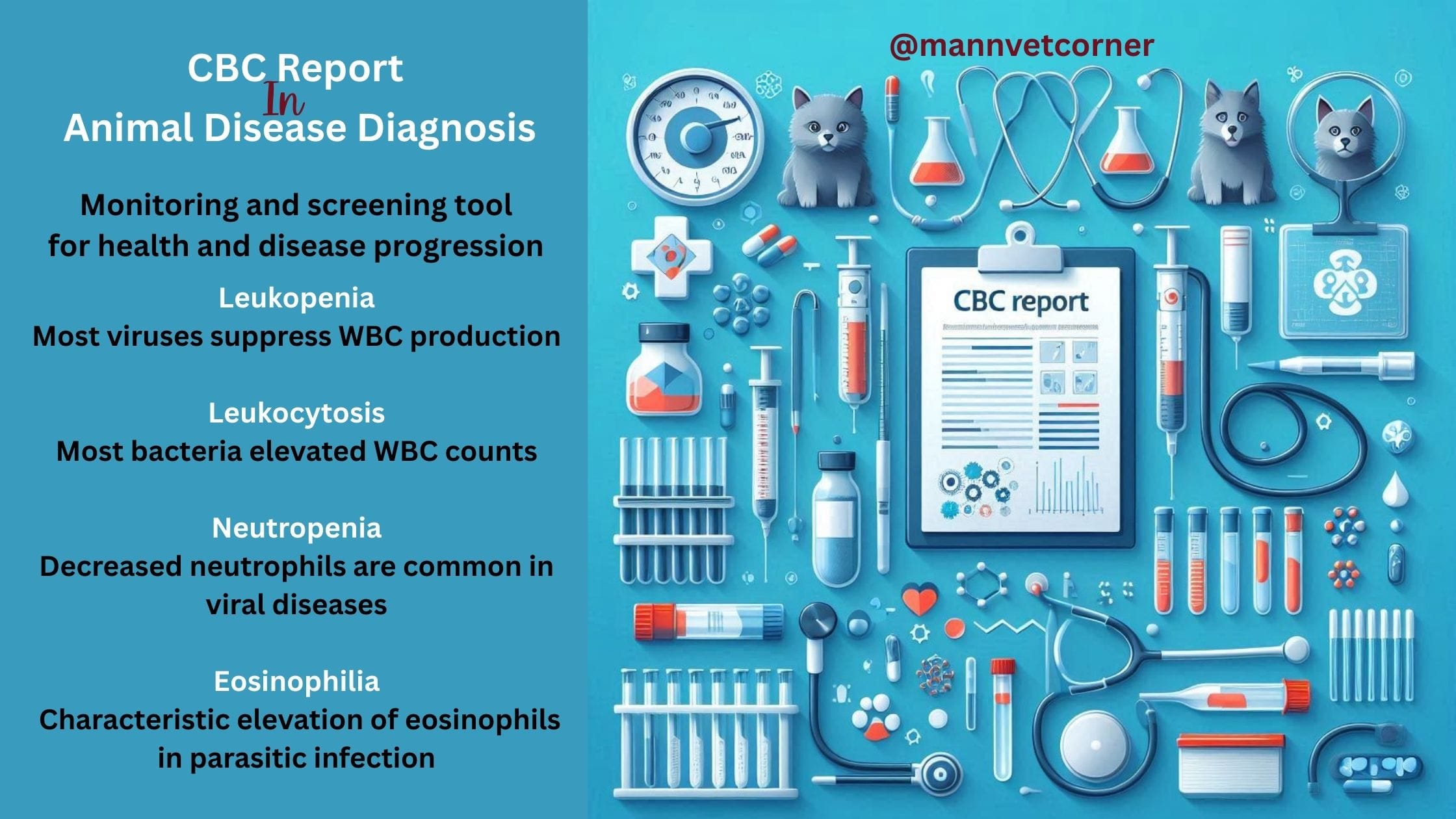
- Leukopenia: Most viruses suppress white blood cell production
- Lymphocytosis: Some viral infections may cause increased lymphocytes
- Neutropenia: Decreased neutrophils common in viral diseases
- Thrombocytopenia: Some viruses affect platelet production
Bacterial Infections
- Leukocytosis: Elevated white blood cell counts typical
- Neutrophilia with left shift: Increased neutrophils including immature forms
- Toxic changes: Neutrophils may show morphological abnormalities
- Lymphopenia: Stress-induced decrease in lymphocytes
Parasitic Infections
- Eosinophilia: Characteristic elevation of eosinophils
- Anemia: Blood loss from parasites affects red blood cell parameters
- Variable WBC counts: Total white cells may be normal, increased, or decreased
Immune-Mediated Diseases
- Regenerative anemia with spherocytes: Hallmark of immune-mediated hemolytic anemia
- Thrombocytopenia: Immune destruction of platelets
- High reticulocyte count: Bone marrow response to hemolysis
Retroviral Infections
- Progressive pancytopenia: Gradual decrease in all blood cell lines
- Bone marrow suppression: Primary effect on blood cell production
- Atypical lymphocytes: May indicate neoplastic transformation
Limitations and Clinical Decision-Making Rules
Important Limitations
Non-specific Findings: Many diseases produce similar CBC patterns. Leukocytosis can occur with any bacterial infection regardless of location or cause.
Early Disease Detection: CBC changes may not appear immediately after disease onset. There may be 6-12 hours delay before changes become apparent.
Stress Effects: Handling and examination stress can significantly affect results, typically causing neutrophilia and lymphopenia.
Species and Age Variations: Normal ranges differ dramatically between species and age groups. Young and adult animals have different baseline values.
Clinical Decision-Making Rules
Never Use CBC Alone: Always correlate with clinical signs, physical examination, patient history, and signalment.
Serial Monitoring: Trends over time provide more diagnostic value than single results.
Pattern Recognition: Look for patterns across multiple parameters rather than focusing on individual abnormalities.
Consider Clinical Context: The same CBC abnormality may have different significance depending on presentation.
When to Pursue Additional Testing
- Unexplained anemia: Blood chemistry, urinalysis, imaging studies
- Persistent leukocytosis: Culture and sensitivity, imaging
- Thrombocytopenia: Coagulation studies, bone marrow evaluation
- Abnormal cell morphology: Flow cytometry, clinical pathologist consultation
Best Practices for CBC Interpretation
Systematic Approach
- Verify Sample Quality: Check for clotting, hemolysis, or collection artifacts
- Assess Overall Patterns: Evaluate CBC as a whole rather than individual parameters
- Use Appropriate Reference Ranges: Apply species-specific and age-appropriate ranges
- Consider Clinical Context: Interpret results with patient’s history and clinical signs
- Evaluate Trends: Compare to previous results when available
Conclusion
CBC remains an indispensable diagnostic tool in veterinary medicine when properly interpreted within clinical context. The case studies demonstrate how characteristic patterns can guide diagnosis and treatment decisions across various species and disease conditions. However, CBC limitations must be recognized, and results should always be integrated with comprehensive clinical evaluation for optimal patient care. Success in CBC interpretation requires systematic analysis, pattern recognition, and understanding of species-specific variations combined with sound clinical judgment.


Very nice website
And very helpfull quiz for learning and improving
Thank you so much for the kind words, Sarmad! I’m really glad you found the website engaging and the quiz helpful for learning.