

In clinical practice, canine anemia presents a puzzle where patient history and laboratory findings must be methodically aligned. Establishing the regenerative capacity of bone marrow is the fundamental first step in categorizing the underlying etiology. By breaking down the diagnostic evaluation into structured stages, clinicians can narrow down broad differential lists with clarity and precision.
Step One: Reticulocyte Quantification
The absolute reticulocyte count serves as the definitive indicator of erythroid response in the marrow. A high absolute count confirms active peripheral loss or destruction, pointing toward hemorrhage or hemolysis. Conversely, a lack of reticulocytes directs diagnostic focus toward central marrow dysfunction or systemic chronic disease.
Step Two: Evaluating Blood Smear Morphology
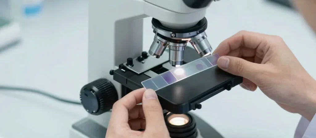
Direct microscopic examination of fresh blood smears reveals cellular features that automated analyzers may overlook. Polychromasia, anisocytosis, and spherocytes offer immediate visual cues pointing toward specific pathological processes like immune-mediated hemolytic anemia. Identifying target cells or Heinz bodies further refines the diagnostic hypothesis regarding oxidative damage.
Step Three: Integrating Clinical Context with Pathology
Laboratory data achieves maximum value only when interpreted alongside physical examination findings such as mucosal pallor or splenomegaly. Developing a patient-specific algorithm prevents missed diagnoses and guides appropriate baseline therapeutic strategies. Systematic follow-up testing ensures early detection of changing bone marrow dynamics.

