

Quick Answer:
What is Lameness in Horses?
| Lameness in horses is an abnormal stance or gait caused by pain or structural problems in the legs, hooves, or body. Common signs include head bobbing, uneven steps, weight shifting, behavior changes, and swelling in the legs or hooves. Causes range from hoof abscesses and laminitis to arthritis, tendon injuries, and navicular syndrome. Diagnosis requires veterinary evaluation using physical exams and movement assessment, followed by treatment including rest, special shoeing, anti-inflammatory medicine, and rehabilitation. So, let’s dive into the Horse Lameness: Complete Guide to Signs, Causes, Diagnosis & Treatment. |
Introduction
A lousy horse is every equine owner’s concern. Whether you’re managing a performance horse, a recreational mount, or a breeding animal, lameness affects mobility, athletic ability, and quality of life. Detecting lameness early—before it becomes severe—is critical for successful treatment and recovery.
This comprehensive guide covers the signs of horse lameness, common causes, how veterinarians diagnose the problem, and evidence-based treatment and prevention strategies. Whether you’re troubleshooting a recent gait change or building a proactive lameness-prevention program, you’ll find practical, expert-backed information here.
Signs of Lameness in Horses: What to Watch For
The earliest signs of lameness are subtle. Some horses show obvious head bobbing or refusal to move; others display minor gait changes only visible to a trained eye. Recognizing these signals allows you to intervene early.
Common Physical Signs of Lameness
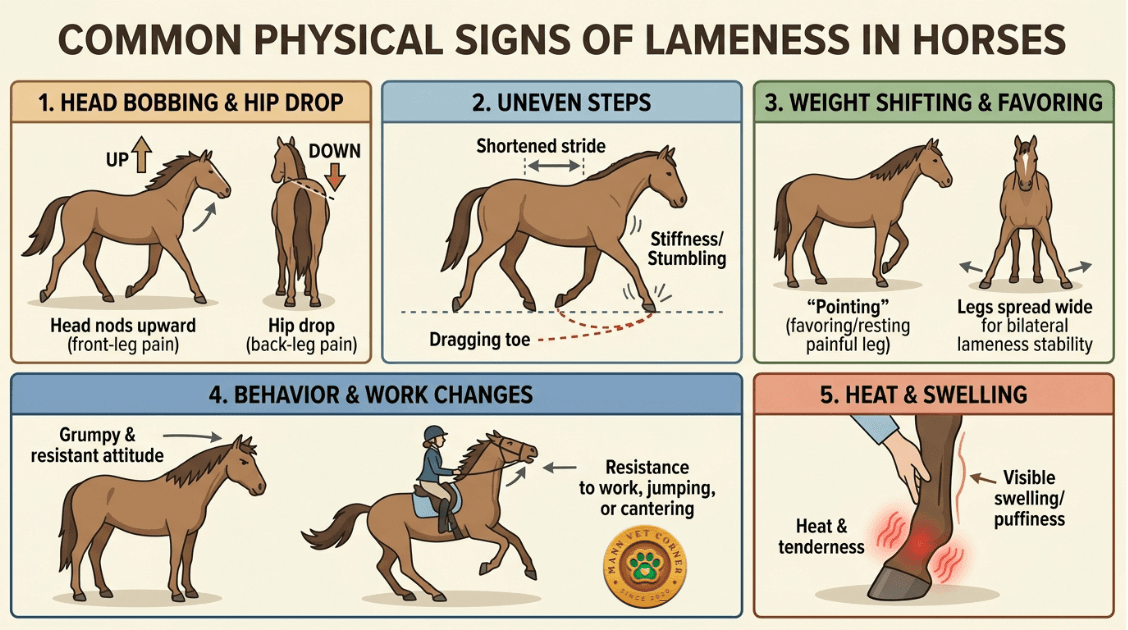
Head Bobbing
A sick horse with front-leg pain often nods its head upward when the painful leg strikes the ground. This reflex shifts weight away from the painful limb. Back-leg lameness may show as a hip drop (the hip on the painful side sinks lower) rather than head bobbing.
Uneven Steps
Shortened stride length, dragging toes, stumbling, or an uneven hip rise in the hindquarters all indicate lameness. The horse may appear stiff, especially after rest or during initial warm-up.
Weight Shifting and Favoring
An uncomfortable horse stands with more weight on the healthy leg. At rest, it may repeatedly rest or pick up the painful limb—a behavior called ‘pointing.’ Horses with bilateral lameness (both front or both hind legs affected) may stand with legs spread wide for stability.
Behavior and Work Changes
Lameness often triggers behavioral shifts: a normally cheerful horse becomes grumpy, resistant to work, reluctant to move in one direction, or unwilling to jump or canter. Some horses become dangerous to ride due to unpredictable movement patterns.
Visible Heat and Swelling
Inflammation and infection cause warm spots and puffy areas on the lower legs or hooves. These may be cold to the touch in chronic cases (like arthritis) but warm and tender in acute injuries.
Lameness Signs by Affected Limb
| Affected Limb | Primary Observable Sign | Severity Indicator |
| Front Left or Right | Head bobs upward when painful leg lands | Obvious head nod = severe; subtle bobbing = mild |
| Hind Left or Right | Hip drop; painful side hip sinks lower | Visible hip asymmetry = clear lameness |
| Multiple Legs (Bilateral) | Stiff, shuffling gait; shortened stride overall | Horse may refuse to move; severe pain |
Common Causes of Lameness in Horses
Lameness stems from pain or structural defects. Understanding the root cause is essential for targeted treatment.
Hoof-Related Causes
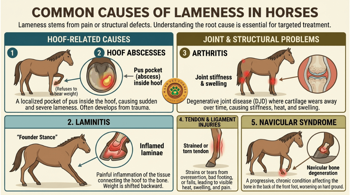
Hoof Abscesses
A hoof abscess is a localized pocket of pus that builds pressure inside the hoof capsule, causing severe, sudden lameness. Abscesses typically develop after hoof trauma, puncture wounds, or infection through a crack. Affected horses often refuse to bear weight on the limb. Drainage and topical treatment usually resolve the abscess in 1–3 weeks.
Laminitis
Laminitis is painful inflammation of the laminae (the tissue connecting the hoof wall to the bone inside). It can be acute (sudden) or chronic. Common triggers include grain overload, endotoxemia (from infection or colic), obesity, and metabolic disorders like insulin resistance. Horses with laminitis show a characteristic ‘founder stance’—shifted weight to the hind legs with the front legs stretched forward.
Joint and Structural Problems
Arthritis
Degenerative joint disease (DJD) or osteoarthritis develops over time as cartilage wears away. Common in older horses and performance animals, arthritis causes stiffness (especially after rest), heat, and swelling. Radiographs reveal bone changes. Management includes rest, joint injections, anti-inflammatory medicine, and joint supplements.
Tendon and Ligament Injuries
High-impact exercise, overexertion, bad footing, or falls can strain or tear the tendons and ligaments that support the leg. Soft-tissue injuries range from mild (heat and swelling) to severe (complete tears requiring months of rehabilitation). Ultrasound helps assess injury severity.
Navicular Syndrome
This chronic, degenerative condition affects the navicular bone and surrounding structures in the back of the foot. It typically causes front-leg lameness that worsens on hard ground and improves with work initially (“warming out” of lameness). Navicular syndrome is progressive and requires long-term management.
Comparison: Common Lameness Causes
| Cause | Onset | Key Symptoms | Duration |
| Hoof Abscess | Sudden (overnight) | Severe pain; foot heat; reluctance to move | 1–3 weeks with drainage |
| Laminitis | Acute or gradual | “Founder stance”; digital pulse; hoof heat | Weeks to months (chronic) |
| Arthritis | Gradual | Stiffness after rest; swollen joints | Chronic (years) |
| Tendon/Ligament Injury | Acute (during work) | Heat; swelling; sudden lameness | Weeks to months (recovery) |
| Navicular Syndrome | Gradual | Worsens on hard ground; improves with work | Progressive (long-term) |
How Veterinarians Diagnose Horse Lameness
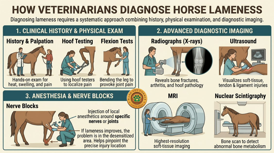
Diagnosing lameness requires a systematic approach combining history, physical examination, and diagnostic imaging.
Clinical History and Physical Exam
Your veterinarian begins by asking questions: When did you first notice the lameness? Did it start suddenly or gradually? Has the horse had prior injuries? Any recent changes in work, shoeing, or footing? A thorough physical examination includes:
Palpation (hands-on examination) for heat, swelling, and pain sensitivity
Hoof testing with hoof testers to localize pain in the foot
Flexion tests that bend the leg to provoke pain from joints or soft tissues
Observation of the horse at walk, trot, and canter on hard and soft surfaces
Advanced Diagnostic Imaging
When physical findings are unclear, imaging provides detailed visualization:
Radiographs (X-rays): Reveal bone fractures, arthritis, navicular changes, and hoof pathology
Ultrasound: Visualizes soft-tissue injuries in tendons, ligaments, and joint structures
MRI (Magnetic Resonance Imaging): Provides highest-resolution soft-tissue imaging for complex cases
Nuclear Scintigraphy (bone scan): Detects areas of abnormal bone metabolism
Anesthesia and Nerve Blocks
Local anesthetics are injected around specific nerves or joints. If lameness improves after a block, the problem lies in the desensitized area. Blocks help pinpoint injury location precisely—critical for managing performance horses with valuable careers at stake.
Treatment and Management of Equine Lameness
Lameness treatment is individualized based on cause, severity, and the horse’s age and intended use.
Rest and Confinement
Box rest (stall confinement) or pasture rest reduces stress on injured structures. Duration varies: soft-tissue injuries may require 6–12 weeks; hoof problems often resolve faster. Limited movement under controlled conditions (handwalking) gradually rebuilds tissue strength during the recovery phase.
Specialized Shoeing and Hoof Care
A skilled farrier plays a crucial role in lameness management. Therapeutic shoeing can:
Relieve pressure on painful areas (e.g., rocker shoes for laminitis)
Improve breakover (the ease with which the toe leaves the ground) to reduce strain
Correct conformational imbalances that contribute to lameness
Trim schedules are typically every 6–8 weeks to maintain optimal hoof balance.
Medications and Injections
Nonsteroidal anti-inflammatory drugs (NSAIDs) like phenylbutazone (Bute) and firocoxib reduce pain and inflammation, enabling the horse to move comfortably during recovery.
Intra-articular injections (into joints) containing corticosteroids and hyaluronic acid reduce joint inflammation and can provide months of relief for arthritis.
Platelet-rich plasma (PRP) and stem cell therapies show promise for soft-tissue regeneration but are costly and require further research.
Rehabilitation and Conditioning
Gradual return to work is essential. After initial rest, handwalking on firm footing progresses to ridden work as tolerated. Proper conditioning rebuilds tissue strength and flexibility, reducing reinjury risk. Many owners work with equine physical therapists to optimize recovery.
Preventing Lameness: Proactive Hoof and Joint Care
Prevention is always preferable to treatment. Strategic management reduces lameness risk significantly:
Consistent Hoof Care
Schedule farrier visits every 6–8 weeks to maintain hoof balance and remove excess length. Regular trimming prevents contracted heels, clubfoot development, and uneven wear—all risk factors for lameness.
Balanced Nutrition
A diet rich in quality forage, balanced minerals (especially calcium, phosphorus, and trace minerals), and adequate protein supports hoof and joint health. Obesity and insulin resistance increase laminitis risk; weight management is critical.
Appropriate Workload and Footing
Avoid overwork on hard ground, which strains tendons and joints. Provide varied footing (arenas with rubber, pasture, and trails). Young horses need gradual training progression; older animals benefit from lighter work.
Joint Supplements
Products containing glucosamine, chondroitin, and hyaluronic acid may support joint cartilage. Evidence is mixed, but many owners report improved performance when used long-term in working horses.
Frequently Asked Questions About Horse Lameness
What’s the difference between lameness and soreness in horses?
Lameness is an observable gait abnormality caused by pain or structural defect—the horse’s movement is visibly altered. Soreness is mild pain without obvious gait changes; the horse may be reluctant or grumpy but moves relatively normally. Soreness can progress to lameness if untreated.
Can lameness resolve on its own without veterinary care?
Minor, self-limiting injuries (like mild bruises) may improve with rest. However, most lameness requires professional diagnosis and treatment. Delaying care risks permanent damage, chronic pain, or secondary injuries from altered movement patterns. Veterinary evaluation is always recommended.
How long does lameness recovery typically take?
Recovery time varies widely by cause. Hoof abscesses resolve in 1–3 weeks; soft-tissue injuries may require 6–12 weeks or longer; arthritis is chronic and managed long-term. Your veterinarian will provide a specific timeline based on diagnosis.
Is lameness in horses painful? Should I worry about my horse’s comfort?
Yes, lameness is painful. The altered gait and behavioral changes reflect discomfort. Prompt diagnosis and pain management—whether through medication, rest, or targeted therapy—improve quality of life and recovery outcomes. Never ignore signs of lameness.
Can a sad horse be ridden or worked during recovery?
It depends on the cause and severity. Acute injuries require complete rest. During later recovery phases, controlled handwalking or light riding may be permitted under veterinary guidance. Working a severely sad horse can worsen injury and cause compensatory lameness in other limbs.
What is a hoof abscess, and why does it cause such severe lameness?
A hoof abscess is an infected pocket of pus trapped inside the hoof capsule. The hard hoof wall cannot expand, so pressure builds painfully. This extreme pain is why horses with abscesses often refuse to move. Drainage through the sole or coronary band relieves pressure and pain rapidly.
Are certain breeds or ages more prone to lameness?
Yes. Heavy draft breeds are prone to joint problems; racehorses and jumping horses experience higher soft-tissue injury rates. Older horses develop arthritis more commonly. Young horses are vulnerable to growth-related problems. Individual conformation and genetics influence lameness susceptibility.
Is there a cure for navicular syndrome?
Navicular syndrome is progressive and cannot be cured, but it can be managed effectively long-term. Treatment focuses on pain relief (NSAIDs, joint injections), appropriate shoeing, controlled exercise, and activity modification. Many horses remain functional and comfortable with proper management.
What role does farrier care play in lameness recovery?
The farrier is central to recovery. Therapeutic shoeing reduces pressure on painful structures, improves biomechanics, and speeds healing. Coordination between your farrier and veterinarian ensures treatment aligns with the diagnosis. Many recoveries depend on skilled hoof care.
Can I prevent laminitis in my horse?
Laminitis prevention focuses on risk management: maintain ideal body weight, limit grain/rich feeds, ensure quality forage, manage dental health, monitor for metabolic issues (like insulin resistance), and avoid sudden diet changes. For high-risk horses, veterinary monitoring and dietary management are critical.
Conclusion: Taking Action on Horse Lameness
Lameness is a complex problem with many possible causes, but early detection and professional management lead to positive outcomes in most cases. By recognizing the signs, seeking prompt veterinary care, and implementing preventive strategies, you can keep your horse sound and comfortable.
Whether your horse is a valued riding companion or a competitive athlete, lameness affects both performance and quality of life. Partner with your veterinarian and farrier, maintain consistent hoof care and nutrition, and provide appropriate work and conditioning. These steps dramatically reduce lameness risk and support long-term health.
Don’t wait for lameness to become severe. Trust your instincts, act on early signs, and prioritize expert care. Your horse will thank you with sound, comfortable movement for years to come.
Sources and Further Reading
MSD Veterinary Manual – Lameness in Horses
American Association of Equine Practitioners (AAEP) – Lameness guidelines and best practices
Equine Veterinary Education Journal – Research on diagnostic imaging and treatment outcomes
American Farriers Association – Standards for therapeutic shoeing and hoof care
