

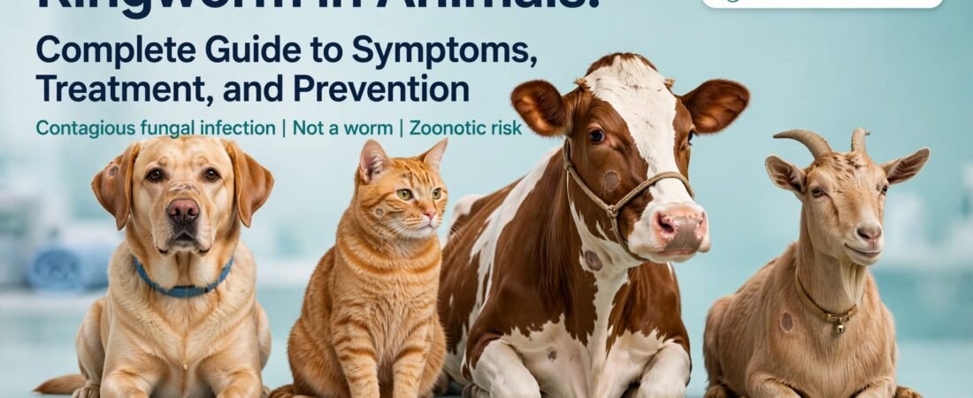
Ringworm is a contagious fungal skin infection caused by dermatophyte fungi, not an actual parasitic worm. It affects pets, livestock, and wildlife, causing circular or patchy hair loss, skin scaling, crusting, and mild-to-absent itching depending on the animal species. The infection spreads through direct contact with infected animals or contaminated environments, and it poses a zoonotic risk to humans. Veterinary diagnosis uses special lighting (Wood’s lamp) or fungal culture testing, and treatment combines prescribed antifungal medications (topical shampoos, ointments, and oral treatments) with thorough environmental disinfection to prevent reinfection and transmission of ringworm (dermatophytosis) in animals
Introduction: Understanding Ringworm in Domestic and Farm Animals
Ringworm remains one of the most common fungal skin infections affecting animals worldwide, with a prevalence estimated between 15-20% in shelters and rescue facilities. Despite its colloquial name, ringworm (dermatophytosis) is not caused by a parasitic worm but by fungal organisms belonging to the genera Microsporum, Trichophyton, and Epidermophyton. These dermatophytes are keratinophilic fungi that colonize the outer layers of skin, hair, and nails, causing visible clinical signs ranging from mild scaling to severe alopecia (hair loss).
The zoonotic nature of ringworm—its ability to transmit from animals to humans—makes it a significant public health concern. Pet owners, veterinarians, and livestock handlers face particular risk of infection through direct contact with infected animals or contaminated fomites (inanimate objects). Understanding the etiology, transmission dynamics, clinical presentation, diagnostic protocols, evidence-based treatment strategies, and prevention measures is essential for effective management and control.
What Causes Ringworm in Animals? The Fungal Pathogens Behind Dermatophytosis
Ringworm is caused by dermatophyte fungi, which are specialized organisms that preferentially colonize keratinized tissues (skin, hair, nails). These fungi produce keratinolytic enzymes that allow them to break down keratin, the structural protein in hair and skin. The three primary genera responsible for dermatophytosis in animals are:
Microsporum species:
Microsporum canis is the most common cause of ringworm in cats and dogs globally, accounting for approximately 50-70% of canine and feline dermatophytosis cases. M. audouinii affects primates and occasionally humans. M. gypseum is a geophilic fungus (soil-dwelling) that affects animals in contact with contaminated soil.
Trichophyton species:
- mentagrophytes is primarily zoophilic (animal-adapted) and affects rodents, rabbits, horses, and occasionally dogs. T. verrucosum affects cattle and is a common cause of ringworm in livestock. T. equinum is specific to equines and is endemic in certain geographic regions.
Epidermophyton species:
- floccosum is primarily a human pathogen but occasionally affects animals through prolonged contact with infected humans.
Ringworm Symptoms in Animals: Recognizing Clinical Signs
Clinical manifestations of dermatophytosis vary by host species, age, immune status, and fungal species involved. Common symptoms include:
Hair Loss (Alopecia):
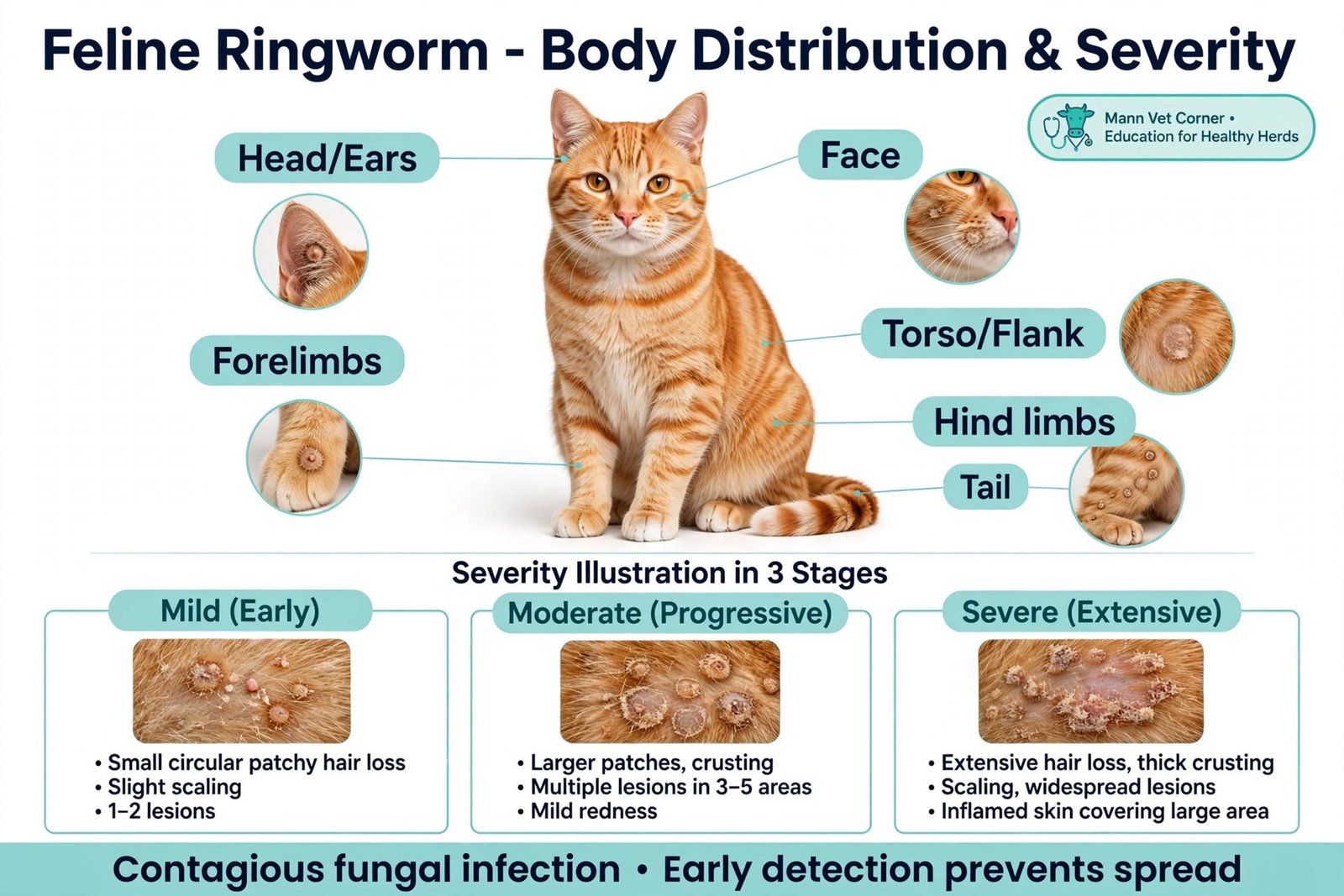
Circular or patchy alopecia is the hallmark clinical sign. Lesions typically appear on the face, ears, limbs, and dorsal surface of the animal. In cats, lesions often begin on the ears, face, and front limbs. In dogs, lesions are more variable but commonly affect the face, ears, and forelimbs. The pattern of hair loss follows fungal growth, which is typically centripetal (expanding outward from a central point).
Skin Changes:
Affected skin exhibits scaling (resembling dandruff), crusting, erythema (redness), and variable degrees of inflammation. Some lesions may appear as gray, powdery scaling on hair shafts. Secondary pyoderma (bacterial infection) can develop, particularly in animals with compromised skin barriers or altered immune function.
Hair Texture Changes:
Hair becomes brittle, breaks easily, and loses normal luster. Affected hairs may pull out painlessly when gently tugged.
Pruritus (Itching):
Contrary to common belief, ringworm typically causes mild-to-absent itching in animals, particularly in cats. When pruritus is severe, it may indicate secondary bacterial infection or concurrent allergic conditions. Dogs may show variable pruritus levels.
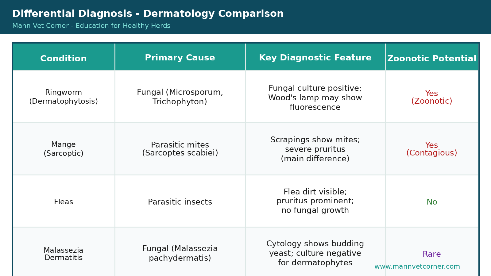
Ringworm vs. Other Fungal and Parasitic Conditions: Diagnostic Differentiation
How Ringworm Spreads: Transmission Routes and Environmental Persistence
Understanding transmission dynamics is critical for implementing effective control measures. Ringworm spreads through multiple routes:
Direct Contact with Infected Animals:
Direct skin-to-skin or hair-to-skin contact with infected animals is the primary transmission route. Fungal spores and mycelial fragments are shed from lesions and adhere to hair, skin, and saliva. Grooming contact, playing, and sleeping in close proximity facilitate transmission.
Contaminated Fomites (Environmental Surfaces):
Dermatophyte spores are highly resistant and can survive on inanimate objects for weeks to months depending on environmental conditions (temperature, humidity, light exposure). Common contaminated sources include:
Bedding and blankets (spores can survive 12+ months)
Grooming tools (brushes, combs, clippers)
Furniture, floors, and carpets
Toys and food/water bowls
Zoonotic Transmission (Animal-to-Human):
Microsporum canis, T. mentagrophytes, and T. verrucosum are zoonotic, meaning they readily transmit from animals to humans. Transmission typically occurs through handling infected animals, grooming, or contact with contaminated materials. Risk is highest in veterinarians, animal handlers, pet owners, and immunocompromised individuals (children, aged, and immunosuppressed). In humans, ringworm often appears as a distinct ring-shaped lesion on exposed skin (arms, face, neck), though multiple morphologies are possible.
Ringworm Diagnosis: Veterinary Testing Methods and Protocols
Accurate diagnosis is essential for initiating appropriate treatment and implementing quarantine measures. Veterinarians employ multiple diagnostic techniques:
Wood’s Lamp (Ultraviolet) Examination:
A Wood’s lamp emits long-wave ultraviolet light (365 nm). Approximately 50-60% of Microsporum canis infections exhibit a distinctive blue-green fluorescence under Wood’s lamp examination. Important caveat: not all ringworm cases fluoresce, and other conditions (lint, soap residue, bacterial biofilms) may show false-positive fluorescence. Therefore, Wood’s lamp examination alone is not sufficient for definitive diagnosis and must be paired with fungal culture or microscopy.
Fungal Culture (Definitive Diagnosis):
Fungal culture is the gold standard for ringworm diagnosis. Samples are collected by brushing the affected area with a sterile brush or by extracting broken hairs and scales. Samples are plated on dermatophyte test medium (DTM) or Sabouraud’s dextrose agar (SDA). Microsporum species typically show growth within 1-3 weeks and produce characteristic color changes (red or pink coloration on DTM). Culture allows for species identification and, when needed, antifungal susceptibility testing. Turnaround time is typically 7-21 days, depending on laboratory procedures.
Microscopic Examination (KOH Preparation):
Hair and scale samples can be treated with potassium hydroxide (KOH), which dissolves cellular material, allowing visualization of fungal spores and hyphae under microscopy. This method provides faster results than culture (24-48 hours) but is less sensitive and specific. KOH microscopy is useful for rapid preliminary assessment but should not replace fungal culture for definitive diagnosis.
Ringworm Treatment: Multimodal Approach to Antifungal Therapy
Effective ringworm management requires a comprehensive approach combining topical medications, systemic antifungals, and environmental decontamination. Treatment protocols are tailored based on species, lesion extent, animal age, and immune status.
Topical Antifungal Therapy:
Prescribed medicated shampoos and ointments are essential components of ringworm treatment. Commonly used agents include:
Chlorhexidine (2-4%) shampoos: Provide broad-spectrum antimicrobial activity and improve skin barrier function. Particularly effective when combined with antifungal agents.
Miconazole (2%) shampoos: Imidazole antifungals that interfere with ergosterol synthesis in fungal cell membranes.
Enilconazole ointments: Topical imidazole with strong antifungal efficacy for localized lesions.
Lime sulfur dips (2-3%): Traditional, sulfur-based antimicrobial that remains effective against dermatophytes; also has scabicidal and antibacterial properties.
Protocol: Most veterinarians recommend twice-weekly medicated shampoo baths for 6-12 weeks, with lathering for 10 minutes to ensure contact time. Topical ointments may be applied to focal lesions between shampoo sessions.
Oral (Systemic) Antifungal Medications:
Oral antifungals are indicated for extensive infections, high-risk animals, or cases not responding to topical therapy alone. First-line oral agents include:
Terbinafine (allylamines class): Fungicidal agent with strong efficacy against dermatophytes. Achieves high concentrations in skin and nail tissue. Typical dose: 10-15 mg/kg PO once daily for 4-6 weeks.
Itraconazole (triazoles class): Azole antifungal with broad spectrum activity. Typical dose: 5-10 mg/kg PO once daily for 4-8 weeks. Often used in alternating pulse therapy (7 days on, 7 days off).
Fluconazole (triazoles class): Azole with good bioavailability. Typical dose: 5-10 mg/kg PO once daily. May be used in combination therapies.
Environmental Decontamination and Quarantine:
Environmental control is often overlooked but is critical to prevent reinfection and transmission. Effective decontamination includes:
Bedding: Wash all bedding, blankets, and fabric toys in hot water (>54°C) with antifungal detergents weekly during treatment and for 2-4 weeks post-recovery.
Grooming tools: Disinfect or replace brushes, combs, and clippers using 1:10 bleach solution or commercial disinfectants (e.g., quaternary ammonium compounds).
Hard surfaces: Vacuum carpets and floors thoroughly, then disinfect with 1:100 diluted household bleach or commercial antifungal sprays. Repeat weekly during treatment.
Furniture: Cover or disinfect furniture where the infected animal rests. Consider removing porous furniture if severely contaminated.
Air filtration: Use HEPA filters to reduce airborne spores in multi-pet households.
Isolation: Ideally, infected animals should be isolated from other pets and immunocompromised household members for 2-3 weeks after treatment initiation, or until clinical improvement is evident.
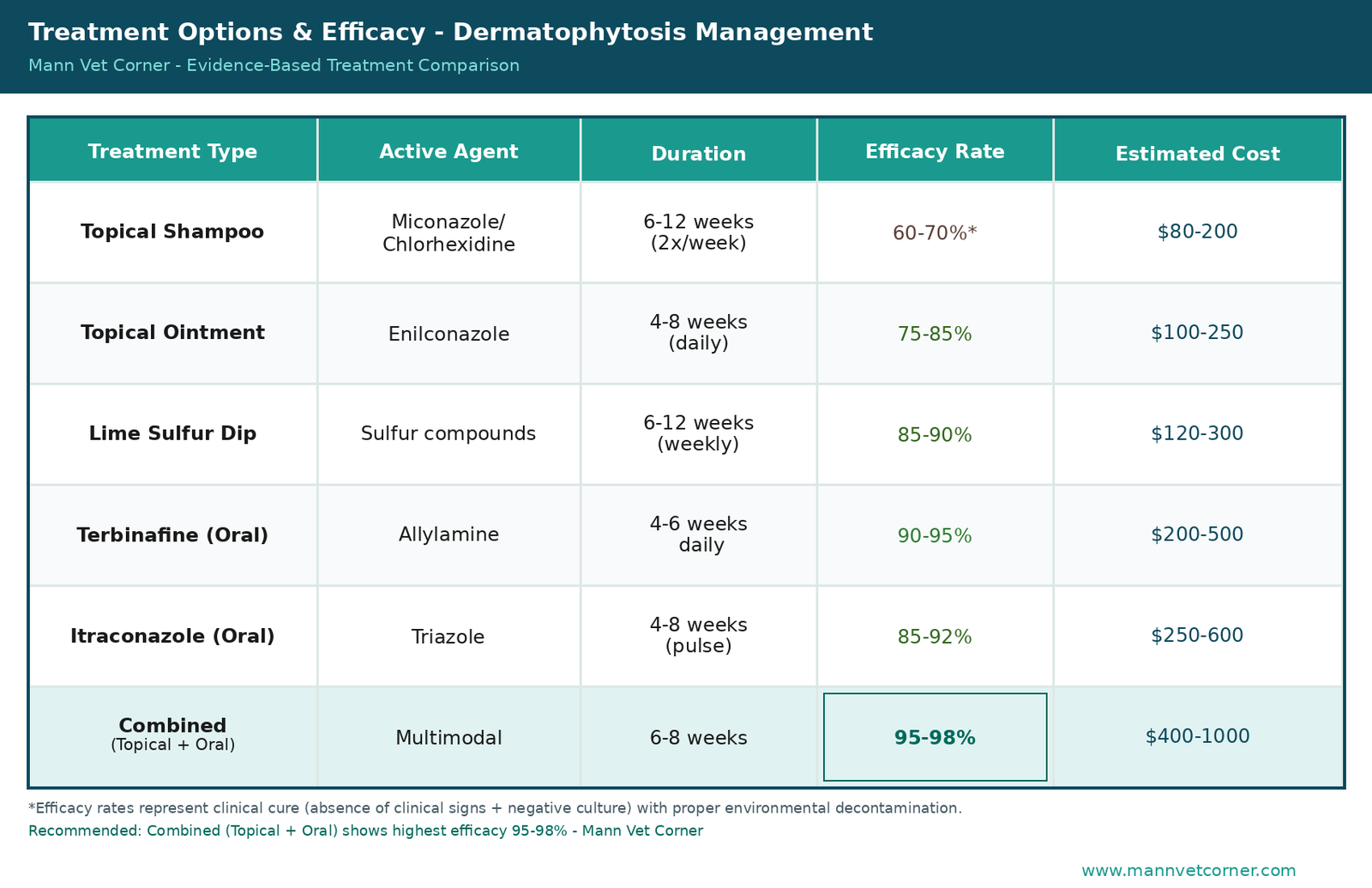
Antifungal Treatment Options: Efficacy and Cost Comparison
Preventing Ringworm: Proactive Strategies for Pet Owners and Livestock Handlers
Prevention is more cost-effective and practical than treatment. Key prevention strategies include:
Quarantine Protocols:
New animals should be quarantined for 2-3 weeks before integration with existing animals. This period allows for observation of clinical signs and preliminary fungal culture results.
Hygiene and Biosecurity:
Practice rigorous hand hygiene before and after handling animals. Use dedicated grooming tools for each animal. Disinfect equipment and facilities regularly using diluted bleach (1:10 solution) or commercial disinfectants.
Regular Grooming and Inspection:
Regular grooming and visual inspection allow early detection of lesions. Prompt veterinary evaluation of any suspicious skin changes enables early diagnosis and treatment.
Environmental Management:
Maintain clean, dry living environments with good ventilation. Reduce humidity levels (fungal spores proliferate in high-humidity conditions). Control wildlife and rodent populations, which may harbor dermatophytes.
Immunocompromised Animal Considerations:
Animals with immunocompromise (young, aged, chronic illness, corticosteroid therapy) are at higher risk for ringworm. Particularly attentive prevention measures and prompter veterinary intervention are warranted.
Zoonotic Ringworm: Human Health Implications and Protective Measures
Ringworm transmission from animals to humans represents a significant public health concern, particularly in veterinary clinics, shelters, farms, and households with young children or immunocompromised individuals.
Most Common Zoonotic Species:
Microsporum canis: Accounts for approximately 70% of zoonotic ringworm cases from pets. Transmission is particularly common from kittens and puppies.
Trichophyton mentagrophytes: Historically associated with rabbit and rodent contact, increasingly identified in companion animal and livestock transmission.
Trichophyton verrucosum: Primarily cattle-associated; risk is highest for farmers and livestock handlers.
Clinical Presentation in Humans:
Human ringworm typically presents as annular (ring-shaped) lesions with central clearing, scale, and erythema on exposed skin (arms, face, neck, hands). However, morphologies vary based on site and individual immune response. Lesions are typically itchy and may persist for weeks without treatment.
High-Risk Populations:
Children (higher transmission risk due to frequent hand-to-face contact and less rigorous hygiene)
Veterinarians and animal health technicians
Shelter and rescue workers
Farmers and livestock handlers
Immunocompromised individuals (HIV/AIDS, organ transplant recipients, chronic corticosteroid users)
Protective Measures for Humans:
Frequent hand washing with soap and water after animal handling
Avoid touching infected animal lesions; use gloves if direct contact is necessary
Maintain personal hygiene and avoid sharing grooming tools or towels
Seek prompt medical evaluation if suspicious skin lesions develop
Frequently Asked Questions About Ringworm in Animals
Q1: Is ringworm actually caused by a worm?
No. Despite the misleading name, ringworm is caused entirely by fungi (dermatophytes), not parasitic worms. The ‘ring’ in ringworm refers to the characteristic ring-shaped appearance of lesions in humans, which results from the pattern of fungal growth and the immune response to it. In animals, lesions often appear as circular to irregular patches of hair loss and scaling rather than distinct rings.
Q2: Can ringworm spread between different animal species?
Yes, though susceptibility varies by species and fungal species. Zoophilic dermatophytes (animal-adapted species like M. canis and T. mentagrophytes) readily transmit between different animal species and to humans. Geophilic species (soil-dwelling) and anthropophilic species (human-adapted) are less commonly transmitted between species. This is why ringworm outbreaks in multi-species households or farms are common if one animal is infected.
Q3: How long does it take for ringworm to develop after exposure?
The incubation period varies but typically ranges from 7-14 days after exposure. Factors influencing incubation include fungal species, inoculum size, skin barrier integrity, and host immune function. Some animals may develop clinical signs within days, while others take 2-3 weeks. This variable incubation period emphasizes the importance of quarantine protocols when introducing new animals.
Q4: Can ringworm be prevented with vaccines?
Ringworm vaccines exist but are not routinely recommended in most countries (including the US and Canada) due to limited efficacy and variable protection. Vaccines appear more beneficial as therapeutic agents (improving clinical recovery) in actively infected animals rather than preventive agents. Standard prevention relies on biosecurity measures, quarantine, and hygiene practices.
Q5: What is the prognosis for ringworm in animals?
Prognosis is generally excellent with appropriate treatment. Most animals achieve clinical cure (resolution of lesions and negative fungal culture) within 6-12 weeks of comprehensive multimodal therapy (topical + oral antifungals + environmental decontamination). Factors affecting recovery include age (younger animals recover faster), immune status, lesion extent, and consistency with treatment protocols. Some animals, particularly immunocompromised individuals, may require extended treatment.
Q6: Can an animal become reinfected after successful treatment?
Yes, reinfection is possible if environmental decontamination is inadequate or if the animal is reexposed to a contaminated environment. This is why thorough cleaning of bedding, toys, furniture, and floors is critical. Reinfection rates increase significantly if environmental control measures are neglected, emphasizing that treatment is not solely about the animal but also about eliminating environmental spore reservoirs.
Q7: Is it safe to handle an infected animal without gloves?
While casual contact with an infected animal does not guarantee human transmission, direct contact with lesions increases risk, particularly for individuals with compromised skin barriers, young children, and immunocompromised persons. Gloves should be worn when handling infected animals or their bedding. Even without gloves, thorough hand washing immediately after handling significantly reduces transmission risk.
Q8: How long do ringworm spores survive on surfaces?
Dermatophyte spores are remarkably resilient. Survival duration varies by fungal species, surface type, temperature, humidity, and UV exposure. Generally, spores can survive on contaminated fabrics for 12+ months, on hair and debris for several months, and on hard surfaces for weeks to months. This extended environmental persistence underscores why repeated environmental decontamination over weeks-to-months is necessary during and after treatment.
Q9: Do all animals develop clinical signs when infected?
No. Some animals, particularly older animals and those with robust immune function, may harbor dermatophytes without developing clinical lesions. These ‘asymptomatic carriers’ can still shed spores and transmit infection to other animals. Conversely, very young animals, aged animals, and those with immunocompromise are more likely to develop clinical signs. This is why fungal culture screening is recommended for potentially exposed animals even if clinical signs are absent.
Conclusion: Comprehensive Ringworm Management and Future Directions
Ringworm (dermatophytosis) represents a significant challenge in companion animal medicine, livestock management, and public health. While the colloquial name is misleading—the condition is caused by fungi, not parasitic worms—the complexity of transmission dynamics, environmental persistence, and zoonotic potential demands comprehensive understanding and management strategies.
Successful treatment requires a multimodal approach combining topical antifungal therapy, systemic antimycotic agents (when indicated), and rigorous environmental decontamination. Early diagnosis through fungal culture is critical to initiate appropriate treatment promptly and implement quarantine measures to prevent transmission. The zoonotic potential of ringworm—particularly species such as M. canis and T. mentagrophytes—emphasizes the need for protective measures in veterinary clinics, shelters, and households with vulnerable populations.
Prevention through quarantine protocols, biosecurity measures, environmental hygiene, and regular health monitoring remains the most cost-effective and practical approach to ringworm management. Understanding that dermatophyte spores can survive on surfaces for months to years underscores the importance of comprehensive environmental decontamination even after clinical cure is achieved.

